Abstract
May-Thurner syndrome (MTS) traditionally refers to compression of the left common iliac vein between the right common iliac artery and the vertebral body. However, iliac vein compression is common in asymptomatic individuals, and the contemporary concept of nonthrombotic iliac vein lesions (NIVLs) emphasizes that anatomic compression alone does not constitute an indication for treatment. Successful endovascular therapy depends on appropriate patient selection and accurate assessment of clinically significant venous outflow obstruction. Intravascular ultrasound is valuable for lesion characterization, assessment of stenosis severity, stent sizing, selection of landing zones, and confirmation of final stent expansion. Durable reconstruction requires complete coverage of the diseased segment, appropriate stent sizing and expansion, adequate proximal outflow with minimal unnecessary coverage of the contralateral common iliac vein ostium, and adequate distal inflow. In post-thrombotic obstruction, reconstruction may need to extend across the inguinal ligament into the common femoral vein when required to reach a satisfactory inflow segment. This review provides a practical framework for the evaluation and endovascular management of MTS and NIVLs, with emphasis on procedural decision-making relevant to interventional radiologists.
-
Keywords: May-Thurner syndrome; Nonthrombotic iliac vein lesion; Iliac vein stenting; Intravascular ultrasound; Deep vein thrombosis
Introduction
May-Thurner syndrome (MTS), classically characterized by compression of the left common iliac vein (CIV) between the overlying right common iliac artery and the lumbar vertebral body, has long been recognized as a predisposing factor for iliofemoral deep vein thrombosis (DVT) and chronic venous outflow obstruction. In 1957, May and Thurner [
1] described intraluminal venous spurs and proposed that chronic arterial pulsation against the compressed vein caused intimal proliferation and progressive luminal narrowing. Cockett and Thomas [
2] subsequently correlated this anatomic abnormality with clinical manifestations of chronic venous insufficiency and iliofemoral thrombosis.
The contemporary concept of nonthrombotic iliac vein lesions (NIVLs) encompasses a broader spectrum of extrinsic iliac venous compression and intrinsic nonthrombotic lesions [
3-
5]. Importantly, substantial iliac vein compression is frequently observed in asymptomatic individuals [
6,
7]. Thus, anatomic compression on CT alone does not establish clinically significant MTS and should not constitute an indication for stent placement [
3,
6,
7].
Clinical manifestations attributable to iliac venous outflow obstruction include persistent unilateral edema, pain, heaviness, venous claudication, skin changes, venous ulceration, and acute iliofemoral DVT [
3,
8,
9]. Endovascular stenting has become the principal method for restoring venous outflow in appropriately selected patients with symptomatic iliofemoral venous obstruction [
10-
13]. Although the primary focus of this review is MTS and NIVL, acute iliofemoral DVT and chronic post-thrombotic obstruction are also discussed because these conditions may coexist with or reveal underlying iliac venous obstruction and require distinct technical considerations during endovascular reconstruction. This review focuses on practical issues relevant to interventional radiologists, including patient selection, assessment of stenosis, the role of intravascular ultrasound (IVUS), stent sizing and landing zones, distal inflow, antithrombotic therapy, and surveillance.
From May-Thurner Anatomy to Clinically Significant NIVL
The classic MTS configuration consists of compression of the left CIV by the right common iliac artery against the vertebral body. Chronic mechanical compression and repetitive arterial pulsation may promote endothelial injury, intimal proliferation, fibrosis, and intraluminal spur formation [
1,
2]. NIVLs may also occur in configurations other than the classic right common iliac artery-left CIV relationship [
3,
4].
The clinically important distinction is between anatomic compression and clinically significant venous outflow obstruction. CT studies demonstrate substantial iliac vein compression in asymptomatic populations [
6,
7,
14]. Anatomic compression becomes clinically relevant when it produces meaningful outflow obstruction that may contribute to venous hypertension, symptoms, collateral formation, or thrombosis. Raju and Neglen [
4] proposed that NIVLs may act as permissive lesions in chronic venous disease rather than as independently sufficient causes of symptoms. Intervention should therefore target clinically relevant venous outflow obstruction rather than imaging abnormality alone.
Patient Selection: Who Should Be Stented?
Appropriate patient selection is a major determinant of clinical success. A phenotype-based practical approach is summarized in
Table 1. The 2024 multidisciplinary consensus statement recommends consideration of NIVL stenting primarily in patients with significant symptoms attributable to venous outflow obstruction after alternative causes have been evaluated [
3]. Typical manifestations include asymmetric edema affecting quality of life, venous claudication, significant pain or heaviness, advanced chronic venous disease, and venous ulceration. Observational studies and a randomized trial have shown improvement in pain, clinical severity, and quality of life after correction of significant iliac venous obstruction [
9,
15,
16].
Potential competing causes, including superficial venous reflux, lymphedema, systemic or medication-related edema, orthopedic disease, and other causes of chronic pain, should be considered before permanent iliac stent placement. Prophylactic treatment of asymptomatic iliac vein compression is not recommended, even when morphologic compression appears marked [
3].
Patients presenting with acute iliofemoral DVT constitute a separate clinical phenotype. Routine endovascular thrombus removal is not indicated for all proximal DVT. In the ATTRACT trial, pharmacomechanical catheter-directed thrombolysis did not reduce the overall incidence of post-thrombotic syndrome, although selected patients with acute iliofemoral DVT may derive symptomatic benefit [
17]. When thrombus-removal therapy is undertaken and significant residual iliac obstruction persists after restoration of flow, stenting of the culprit lesion may be appropriate [
10].
Standardized clinical assessment can facilitate patient selection and longitudinal evaluation. The Venous Clinical Severity Score is useful for grading the severity of chronic venous disease and for assessing change over time, whereas the Clinical-Etiological-Anatomical-Pathophysiological (CEAP) classification provides a standardized description of chronic venous disease. In patients with previous DVT, the Villalta score is commonly used to diagnose and grade post-thrombotic syndrome. Patient-reported instruments such as Venous Insufficiency Epidemiological and Economic Study–Quality of Life/Symptoms (VEINES-QOL/Sym) complement physician-based measures by quantifying venous symptoms and disease-specific quality of life. These tools are particularly useful when the clinical benefit of intervention is assessed beyond anatomic patency alone.
Imaging Evaluation
Duplex ultrasonography is usually the initial examination for lower-extremity venous symptoms. It is effective for assessment of femoropopliteal DVT and venous reflux but has limited ability to directly evaluate the iliac veins. Reduced respiratory phasicity, continuous venous flow, asymmetric common femoral venous waveforms, and collateral formation may suggest proximal obstruction [
18]. A normal lower-extremity duplex examination does not exclude clinically important iliac venous obstruction.
CT venography is useful for defining iliocaval anatomy, the site and configuration of compression, collateral vessels, thrombus extent, and alternative causes of obstruction. However, apparent compression is influenced by hydration, respiratory phase, patient position, and reference-segment selection, while substantial compression is common in asymptomatic individuals [
6,
7,
14]. Magnetic resonance venography can provide similar anatomic information in selected patients. Cross-sectional imaging should therefore be regarded primarily as an anatomic roadmap rather than an independent determinant of the need for stenting.
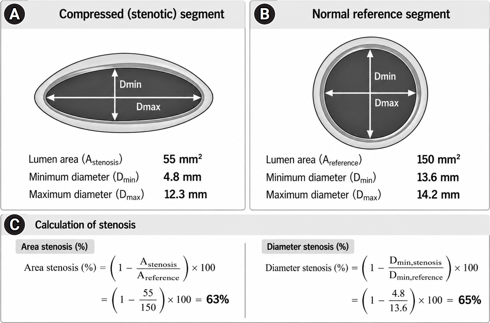
A threshold of 50% stenosis has historically been used to define significant iliac venous obstruction, but no single morphologic threshold reliably distinguishes an asymptomatic anatomic variant from clinically important disease. Stenosis should preferably be assessed by IVUS using the minimum lumen diameter or cross-sectional area at the lesion relative to an adjacent relatively normal reference segment. Because compression frequently produces an eccentric or flattened lumen, direct cross-sectional area measurement may better represent morphologic severity than a single diameter measurement. Diameter and area stenosis are not interchangeable, and the measurement method should be explicitly stated.
The VIDIO trial showed that IVUS-derived measurements were more predictive of clinical improvement after stenting than venographic estimates [
19]. An IVUS-derived area stenosis greater than approximately 54% was associated with clinical improvement, whereas diameter stenosis greater than approximately 61% was useful in the NIVL subgroup [
19].
Fig. 1 schematically illustrates calculation of IVUS-derived area and diameter stenosis by comparing the minimum lumen area at the lesion with the cross-sectional area of an adjacent reference segment. These values should not be interpreted as absolute indications for stenting; current consensus recommends integrating lesion severity with symptoms and the overall clinical context [
3].
Hemodynamic Significance of NIVL
Current recommendations remain primarily symptom-driven, and prophylactic stenting is not recommended in asymptomatic patients solely on the basis of anatomic compression [
3]. Nevertheless, the distinction between anatomic compression and hemodynamically significant obstruction remains incompletely defined. In theory, significant outflow obstruction may increase upstream venous pressure, promote collateral formation, alter flow distribution, and eventually contribute to chronic venous hypertension, edema, skin changes, or thrombosis. This raises the possibility of a continuum from anatomic NIVL to hemodynamically significant NIVL and ultimately to clinically significant disease.
The clinical value of venous pressure measurements, however, remains uncertain. Unlike the arterial circulation, the venous system has high capacitance and extensive collateral pathways. Resting pressure gradients may therefore remain small despite substantial morphologic obstruction and are influenced by patient position, respiration, hydration, collateral circulation, and measurement technique. No validated trans-stenotic pressure-gradient threshold has been established for defining clinically significant NIVL or predicting subsequent deterioration [
3,
10].
Accordingly, elevated venous pressure or a pressure gradient alone should not currently be considered an indication for prophylactic stenting. At the same time, the absence of a current treatment indication does not establish that subclinical venous hypertension is biologically irrelevant. Patients with marked obstruction, extensive collateral formation, abnormal flow patterns, or reproducible hemodynamic abnormalities may represent a distinct higher-risk phenotype. Prospective natural-history studies integrating IVUS-derived stenosis, collateral burden, flow parameters, and resting or exercise-induced pressure measurements are needed to determine whether hemodynamic assessment can improve patient selection.
Role of Intravascular Ultrasound
IVUS has become an important component of contemporary iliac vein intervention because conventional venography provides a 2D projection of a 3D lumen and may underestimate eccentric, flattened, or intraluminal lesions [
19,
20]. IVUS directly demonstrates extrinsic compression, intraluminal spurs, post-thrombotic synechiae, fibrosis, and residual thrombus, while permitting measurement of luminal cross-sectional area and reference venous dimensions.
Procedurally, IVUS is particularly useful for defining the true proximal and distal extent of disease, selecting stent diameter and landing zones, and confirming adequate expansion after deployment. These functions are especially relevant when venography appears satisfactory despite persistent focal compression or when the proximal iliocaval confluence is difficult to define. The Society of Interventional Radiology position statement recommends complementary use of multiplanar venography and IVUS for lesion characterization and procedural guidance when available [
10], and international consensus similarly emphasizes adequate invasive imaging and inflow assessment [
21].
IVUS should nevertheless be interpreted in the clinical context. A large percentage reduction in lumen area does not by itself establish that a lesion is responsible for symptoms, particularly in NIVL, where substantial compression may be present in asymptomatic individuals. Its greatest value is therefore not as an isolated diagnostic threshold but as a procedural tool that integrates morphology, reference caliber, lesion extent, and final reconstruction. In settings where IVUS is unavailable because of reimbursement or resource constraints, careful multiplanar venography and cross-sectional imaging remain important for procedural planning, although they cannot fully reproduce the morphologic and quantitative information provided by IVUS.
Practical Technique of Iliac Vein Stenting
Venous access should be selected according to disease extent. Ipsilateral common femoral vein (CFV) access is convenient for isolated NIVL, whereas more distal femoral or popliteal access can be useful when post-thrombotic disease involves the CFV or when the entire inflow tract must be assessed. Ultrasound-guided access is recommended whenever feasible. In chronic occlusions, the operator should confirm that the guidewire traverses the intended venous channel before dilation.
Balloon angioplasty alone is generally insufficient for clinically significant NIVL because persistent external compression and elastic recoil remain after dilation [
11,
22]. Post-thrombotic lesions contain fixed fibrotic narrowing, webs, synechiae, or chronic occlusions and likewise generally require stent scaffolding for durable luminal restoration.
Stent sizing should be based on the expected normal venous caliber rather than the compressed segment. Undersizing may result in inadequate radial support, incomplete apposition, or migration, whereas excessive oversizing may cause pain or unnecessary extension into adjacent normal venous territory. IVUS measurement of an adjacent relatively normal reference vein provides useful patient-specific information [
3,
10].
Venous stents differ in radial force, crush resistance, flexibility, foreshortening, and deployment behavior [
23]. Fixed diameter recommendations should therefore be considered approximate rather than rigid.
The proximal landing zone is a critical technical consideration in left CIV stenting. A stent that terminates too distally may leave part of the compressed CIV untreated, resulting in residual obstruction, proximal stent compression, or restenosis. Conversely, excessive extension into the IVC may unnecessarily cover the contralateral CIV ostium and alter contralateral venous drainage.
Contralateral DVT after iliac stenting has been evaluated in several observational studies [
24-
27]. Although reported incidence varies and causality is influenced by underlying thrombotic risk, excessive caval extension and contralateral ostial coverage have repeatedly raised concern. More recently, Jacob et al. [
28] quantified postoperative stent position and found an increasing risk of contralateral DVT with increasing coverage of the contralateral iliac vein orifice. These data support avoiding complete or excessive ostial coverage when adequate treatment of the proximal lesion can still be achieved.
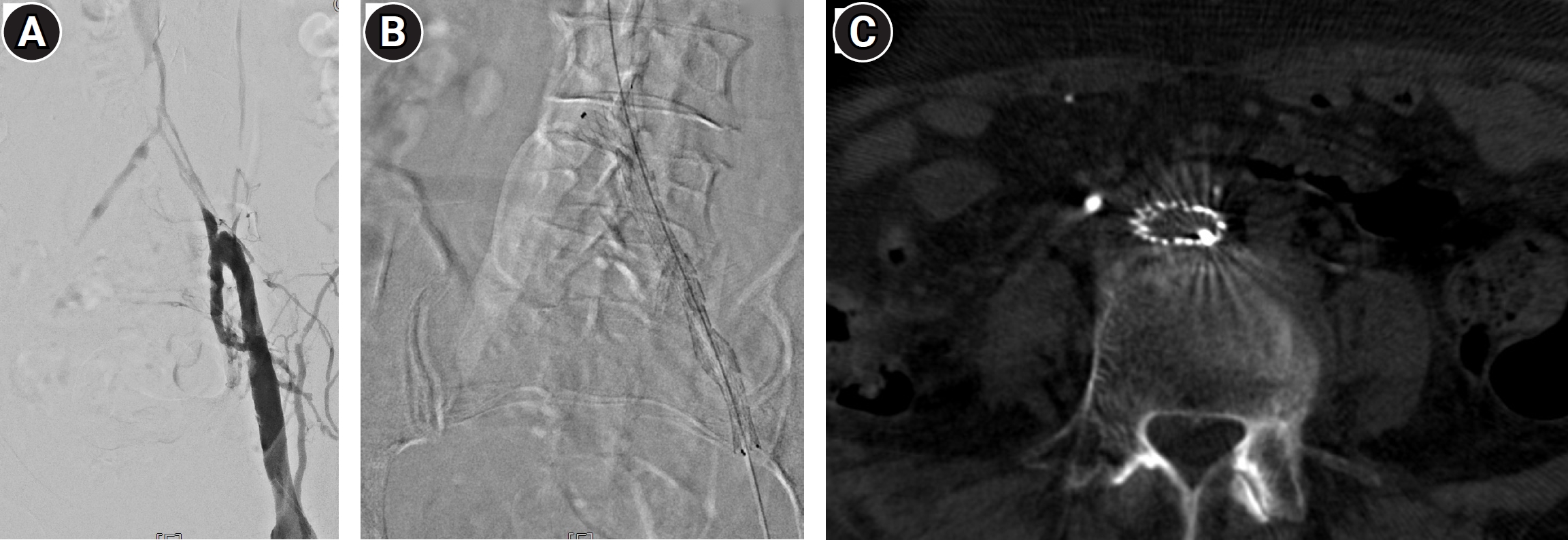
The optimal proximal position should therefore not be defined by a fixed distance of stent extension into the IVC. Instead, the objective is complete coverage of the diseased proximal CIV while preserving as much of the contralateral CIV ostium as possible. Multiplanar venography and IVUS can help define the iliocaval confluence, while roadmap opacification of the contralateral CIV can provide a practical fluoroscopic landmark during deployment (
Fig. 2). Modern dedicated venous stents with more predictable deployment characteristics may further improve landing accuracy.
Failure to control deployment can result in excessive contralateral ostial coverage.
Fig. 3 illustrates unintended stent displacement toward the IVC during deployment with complete coverage of the contralateral CIV ostium and subsequent contralateral iliac thrombosis, emphasizing the importance of deployment behavior and precise positioning at the iliocaval confluence.
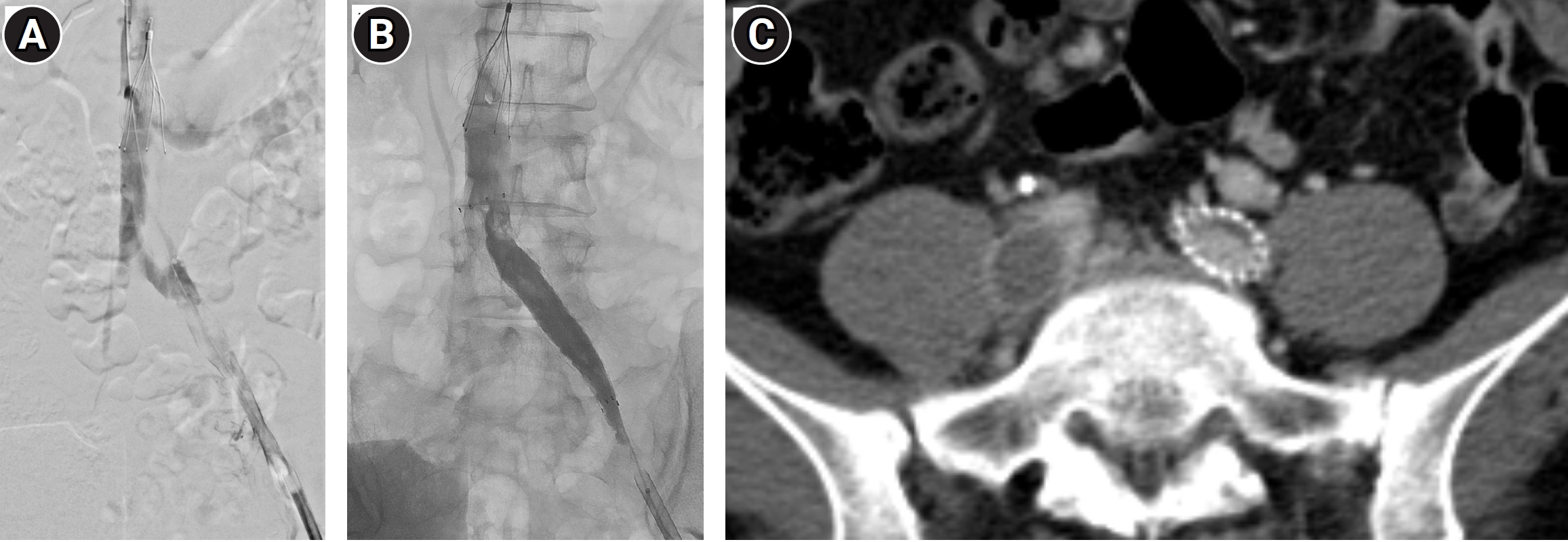
Adequate postdilation is important because a self-expanding stent may remain incompletely expanded at a severe compression site (
Fig. 4).
The distal landing strategy differs between uncomplicated NIVL and post-thrombotic obstruction. In isolated NIVL, the distal landing zone can usually be placed in a relatively normal external iliac vein. In post-thrombotic disease, however, obstruction may extend into the CFV; ending the stent within residual diseased vein may compromise inflow and long-term patency [
10,
21].
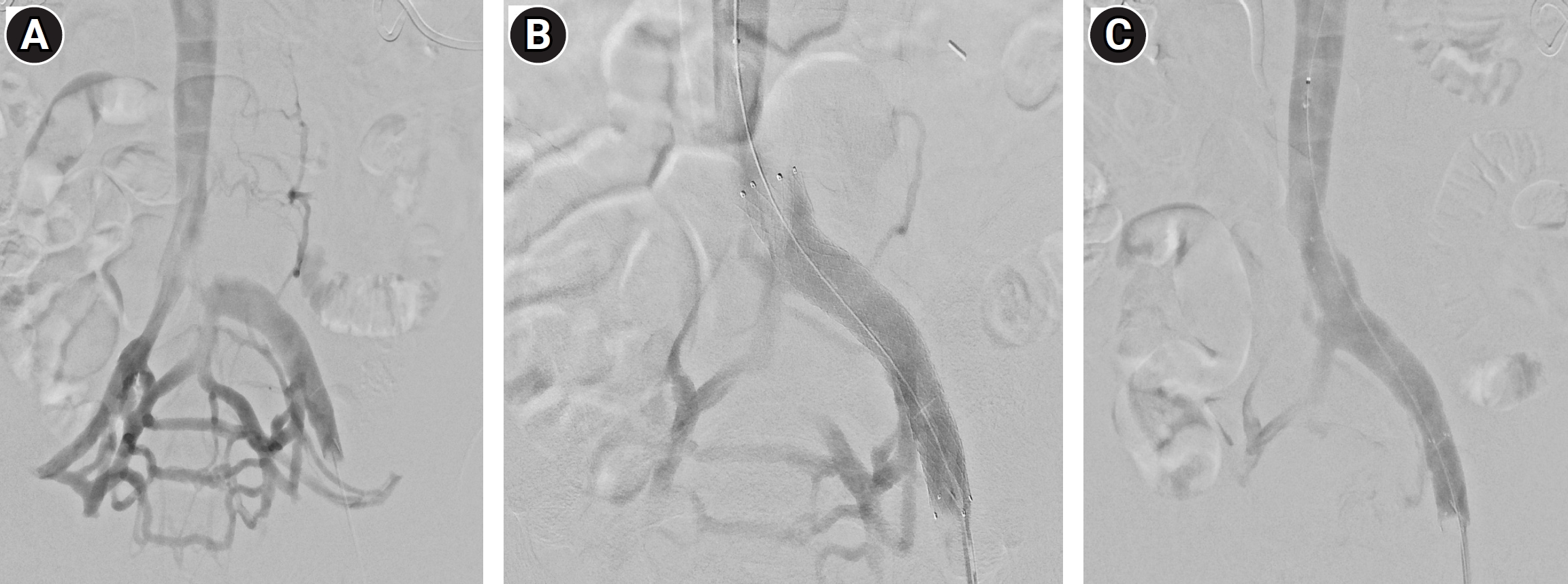
The distal endpoint should therefore be determined by disease extent and inflow quality rather than by the inguinal ligament itself. Available evidence does not support using the inguinal ligament as an absolute distal boundary for venous stenting [
29]. When the femoral vein is chronically occluded, a patent enlarged profunda femoris vein may provide the principal inflow into the reconstructed iliac segment [
30]. Extension into the profunda itself is generally reserved for selected salvage situations [
31].
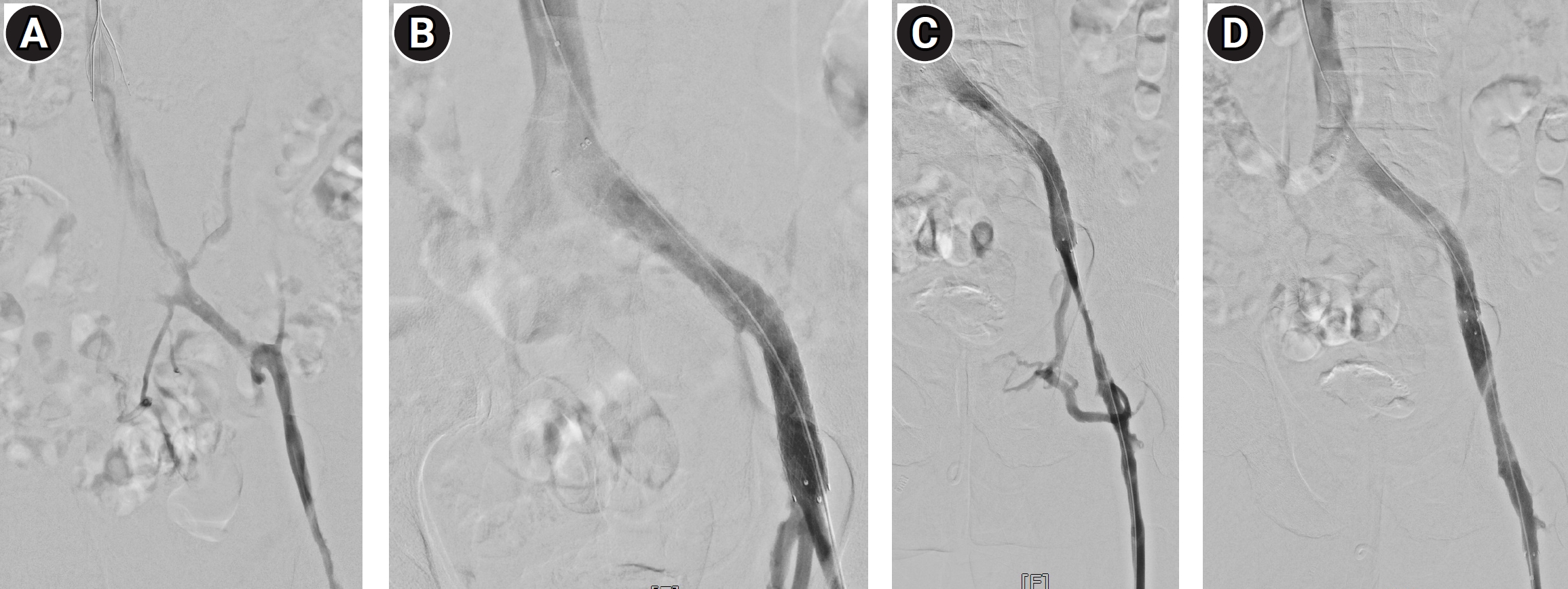
Fig. 5 demonstrates persistent distal post-thrombotic stenosis after proximal iliac stenting, requiring extension into the distal diseased segment to restore adequate inflow.
Acute Iliofemoral DVT and Underlying Iliac Compression
MTS may first become clinically apparent as acute left-sided iliofemoral DVT. Endovascular thrombus removal should not be routinely applied to every proximal DVT; however, carefully selected patients with acute iliofemoral DVT, severe symptoms, good functional status, low bleeding risk, and reasonable life expectancy may benefit from an early thrombus-removal strategy [
17].
After thrombus clearance, the iliac segment should be reassessed for residual thrombus, chronic post-thrombotic abnormalities, and underlying NIVL. IVUS is particularly useful because venography may underestimate residual obstruction [
19,
20]. When a significant culprit lesion persists, stenting is commonly required to maintain venous outflow [
10-
12].
IVC filter placement should not be considered a routine component of endovascular DVT treatment. When used, it should be reserved for selected circumstances in which temporary protection from pulmonary embolization is considered clinically necessary, with retrieval planned when the indication has resolved. In the illustrative cases in
Figs. 3 and
5, the filters were placed prophylactically because of concern for pulmonary thromboembolism during the intervention. This case-specific rationale should not be interpreted as a recommendation for routine filter placement.
Chronic Post-Thrombotic Obstruction
Post-thrombotic iliac obstruction is a more complex phenotype than uncomplicated NIVL. Lesions are often longer and irregular, with synechiae, fibrotic webs, chronic occlusions, and extension into the external iliac or common femoral veins. Recanalization may require more extensive tract preparation, and durable reconstruction is more dependent on adequate inflow. These differences should be recognized when interpreting outcomes across venous stent studies.
Long-term observational studies demonstrate durable clinical benefit in selected patients with chronic iliocaval obstruction [
12,
13,
32], although patency is consistently lower than in NIVL. Across prospective dedicated-stent studies, outcomes have generally been more favorable in nonthrombotic than in post-thrombotic lesions [
33-
37]. The major studies and their practical implications are summarized in
Table 2.
Randomized evidence has also strengthened the rationale for intervention in appropriately selected patients with clinically significant post-thrombotic syndrome. The C-TRACT trial demonstrated greater improvement in post-thrombotic severity and health-related quality of life with an endovascular strategy including iliac stenting compared with standard care alone, although bleeding events were more frequent with intervention [
38]. Thus, potential clinical benefit should be balanced against the greater technical complexity, lower expected patency, and antithrombotic burden of post-thrombotic reconstruction.
Dedicated Venous Stents
Dedicated venous stents were developed to address the biomechanical requirements of the iliofemoral venous system, including radial and crush resistance, flexibility, conformability, and deployment accuracy. Prospective studies of the Venovo, Vici, Abre, and Zilver Vena systems have demonstrated favorable mid- and long-term outcomes [
33-
37]. This section is intended to emphasize device-related considerations rather than to repeat disease-specific outcomes, which are summarized in
Table 2. Because the pivotal studies differed in patient populations, lesion phenotypes, patency definitions, and follow-up methods, their results should not be interpreted as direct head-to-head comparisons. In practice, lesion characteristics and operator familiarity with device behavior remain important considerations.
Antithrombotic Therapy after Iliac Vein Stenting
Postprocedural antithrombotic therapy remains poorly standardized and should be tailored to the underlying disease phenotype. For uncomplicated NIVL without previous thrombosis, the 2024 multidisciplinary consensus does not support mandatory anticoagulation solely because a venous stent has been implanted [
3]. In practice, antithrombotic therapy in this setting may range from no routine therapy to a limited course of antiplatelet or anticoagulant treatment according to patient-specific thrombotic and bleeding risks; comparative evidence is insufficient to establish a single preferred regimen. Earlier international Delphi consensus favored anticoagulation for 6–12 months after venous stenting and low-molecular-weight heparin during the first 2–6 weeks, but no consensus was reached regarding long-term antiplatelet therapy [
39]. These recommendations reflect expert practice patterns rather than high-level comparative evidence.
After stenting performed during treatment of acute DVT, therapeutic anticoagulation should follow established venous thromboembolism principles, considering whether the event was provoked, recurrence history, thrombophilia, ongoing risk factors, and bleeding risk. A finite course is generally appropriate after a single provoked DVT, whereas recurrent or persistent thrombotic risk may justify extended therapy. In chronic post-thrombotic obstruction, long stented segments, poor inflow, recurrent thrombosis, or residual thrombotic disease may favor more intensive or prolonged anticoagulation. The role of adjunctive antiplatelet therapy remains uncertain, and systematic reviews continue to emphasize the lack of comparative evidence [
40]. Thus, the presence of a venous stent alone should not automatically imply lifelong anticoagulation.
Follow-up and Surveillance
The purpose of iliac vein stenting is clinical improvement, not simply normalization of imaging findings. Follow-up should assess edema, pain, venous claudication, skin changes, ulcer healing, functional status, and quality of life together with stent patency. Duplex ultrasonography is commonly used for surveillance, with cross-sectional imaging or repeat venography/IVUS reserved for inconclusive ultrasound or suspected recurrent obstruction.
No universally accepted surveillance interval has been established. Follow-up should therefore be individualized according to disease phenotype, stent extent, inflow quality, thrombotic history, and clinical response, with greater vigilance in post-thrombotic disease because of its higher risk of stent thrombosis [
33-
37].
Complications and Technical Pitfalls
Potential complications include access-site bleeding, venous rupture, stent migration, stent compression, in-stent restenosis, thrombosis, and contralateral DVT. Venous rupture during dilation or stenting is uncommon but may require prolonged balloon tamponade or additional intervention. Stent migration is also uncommon with appropriately sized modern venous stents but can be serious if central embolization occurs; appropriate sizing, sufficient length, and secure landing are therefore essential.
Stent thrombosis is more common in thrombotic and post-thrombotic disease than in uncomplicated NIVL [
33-
37]. When early occlusion occurs, poor inflow, residual thrombus, incomplete lesion coverage, and inadequate stent expansion should be considered. Contralateral DVT deserves particular attention after left CIV stenting, and unnecessary coverage of the contralateral ostium should be avoided when this can be achieved without undertreating the proximal lesion [
24-
28].
The major technical pitfalls can therefore be understood as failures of reconstruction rather than isolated procedural errors. Incomplete lesion coverage leaves residual obstruction; excessive extension sacrifices normal venous territory; undersizing or underexpansion compromises luminal restoration; and an inadequate distal landing zone limits inflow. Durable reconstruction requires that these factors be considered together rather than optimized independently.
Practical Approach for the Interventional Radiologist
A practical strategy begins by determining whether the patient's symptoms are compatible with iliac venous outflow obstruction and by excluding important competing causes. Duplex ultrasonography and cross-sectional imaging are then used to define anatomy and classify the disease as nonthrombotic, acute thrombotic, or chronic post-thrombotic. In patients selected for intervention, multiplanar venography and preferably IVUS should define lesion morphology, severity, reference caliber, and proximal and distal extent. A phenotype-based practical summary is provided in
Table 1.
When stenting is performed, the reconstruction should completely cover the diseased segment with an appropriately sized and adequately expanded stent. Proximally, adequate outflow should be obtained while minimizing unnecessary coverage of the contralateral CIV ostium. Distally, the landing zone should provide reliable inflow; in post-thrombotic disease, this may require extension across the inguinal ligament into the CFV. The appropriate reconstruction therefore depends on disease phenotype rather than on a uniform stenting strategy.
Conclusion
Contemporary management of MTS and NIVLs should focus on clinically significant iliac venous outflow obstruction rather than anatomic compression alone. Careful patient selection is fundamental, and no single morphologic stenosis threshold should be used as an absolute indication for intervention. IVUS complements venography by defining lesion extent, reference venous caliber, stent sizing, landing zones, and final expansion.
Durable iliac venous reconstruction requires complete treatment of the diseased segment while avoiding unnecessary coverage of normal venous anatomy. Proximal coverage should preserve contralateral iliac outflow whenever feasible, and the distal landing zone should provide adequate inflow, particularly in post-thrombotic disease. These principles, applied according to disease phenotype, provide a practical framework for safe and effective iliac vein stenting.
Conflicts of interest
No potential conflict of interest relevant to this article was reported.
Funding
None.
Acknowledgments
None.
Data availability statement
Data sharing does not apply to this article as no datasets were generated or analyzed during the current study.
Fig. 1.Schematic illustration of area and diameter stenosis measurements. (A) Compressed segment showing reduced lumen area and minimum diameter. (B) Normal reference segment. (C) Calculation of area and diameter stenosis using the corresponding measurements from the compressed and reference segments. All measurements are hypothetical and provided for illustrative purposes.
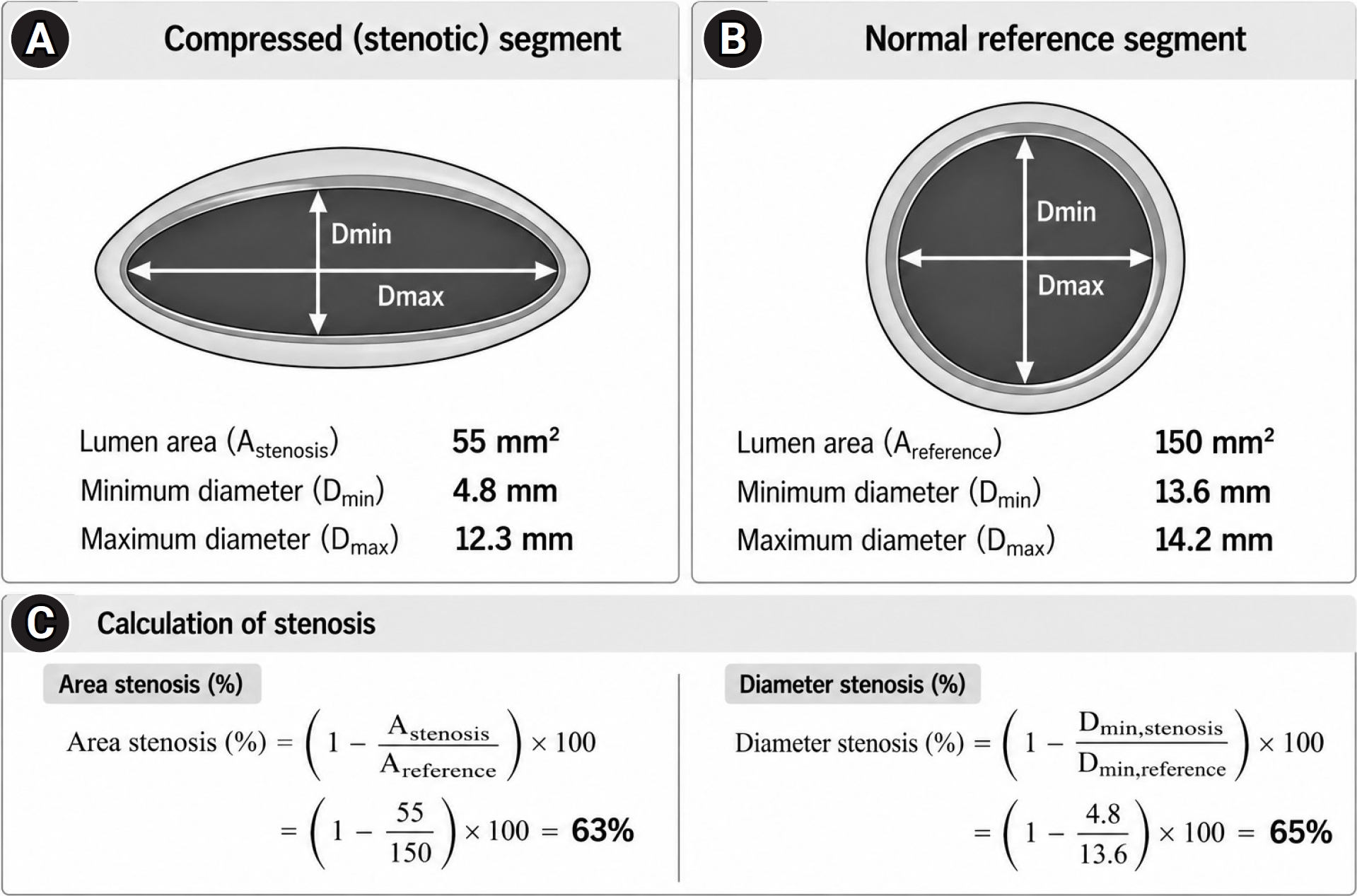
Fig. 2.Iliac vein stenting after catheter-directed thrombolysis in a 74-year-old woman with a 2-week history of left leg swelling. (A) Completion venography after thrombolysis shows near-complete thrombus removal, with severe stenosis of the left common iliac vein and prominent collateral veins. The left external iliac vein appears normal without chronic post-thrombotic change and serves as a distal reference segment. (B) A Cobra catheter positioned in the right common iliac vein is used to delineate the contralateral common iliac vein ostium on roadmap imaging. The stent is deployed to maximize coverage of the stenotic segment while minimizing coverage of the contralateral common iliac vein ostium. (C) Post-stenting cone-beam CT demonstrates stent configuration and expansion.
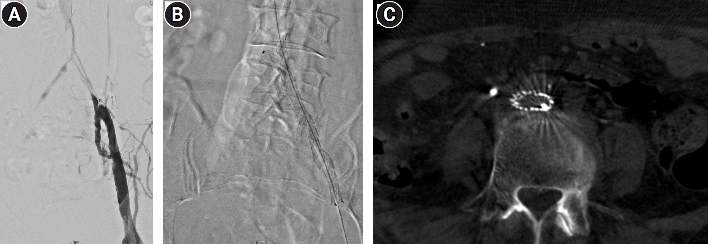
Fig. 3.Contralateral iliac vein thrombosis after iliac vein stenting in a 48-year-old woman presenting with a 5-day history of left leg swelling. (A) Initial venography demonstrates chronic post-thrombotic changes involving the left iliac vein. (B) During stent deployment, unintended stent displacement toward the IVC results in complete coverage of the right common iliac vein ostium. The migration was considered to be related to severe stenosis of the external iliac vein. (C) CT obtained 11 months later because of right leg swelling shows a patent left iliac vein stent but total thrombotic occlusion of the right iliac vein. Venous access was obtained through the left popliteal vein. An IVC filter was placed prophylactically because of concern for pulmonary thromboembolism during the procedure.
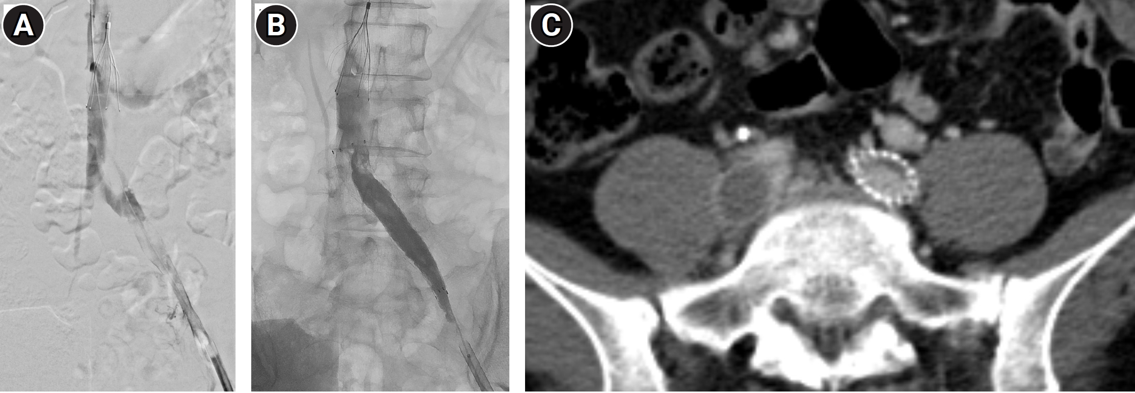
Fig. 4.Inadequate stent expansion requiring additional stent placement in a 59-year-old man with left leg edema after palliative ureterectomy for left ureteral cancer. (A) Venography demonstrates severe stenosis of the left common iliac vein with prominent collateral venous drainage. (B) After initial stent placement and balloon angioplasty, the stent remains incompletely expanded, with persistent collateral venous filling. (C) After placement of a second stent and additional balloon dilatation, the reconstructed iliac vein is widely patent and collateral venous filling has nearly disappeared.
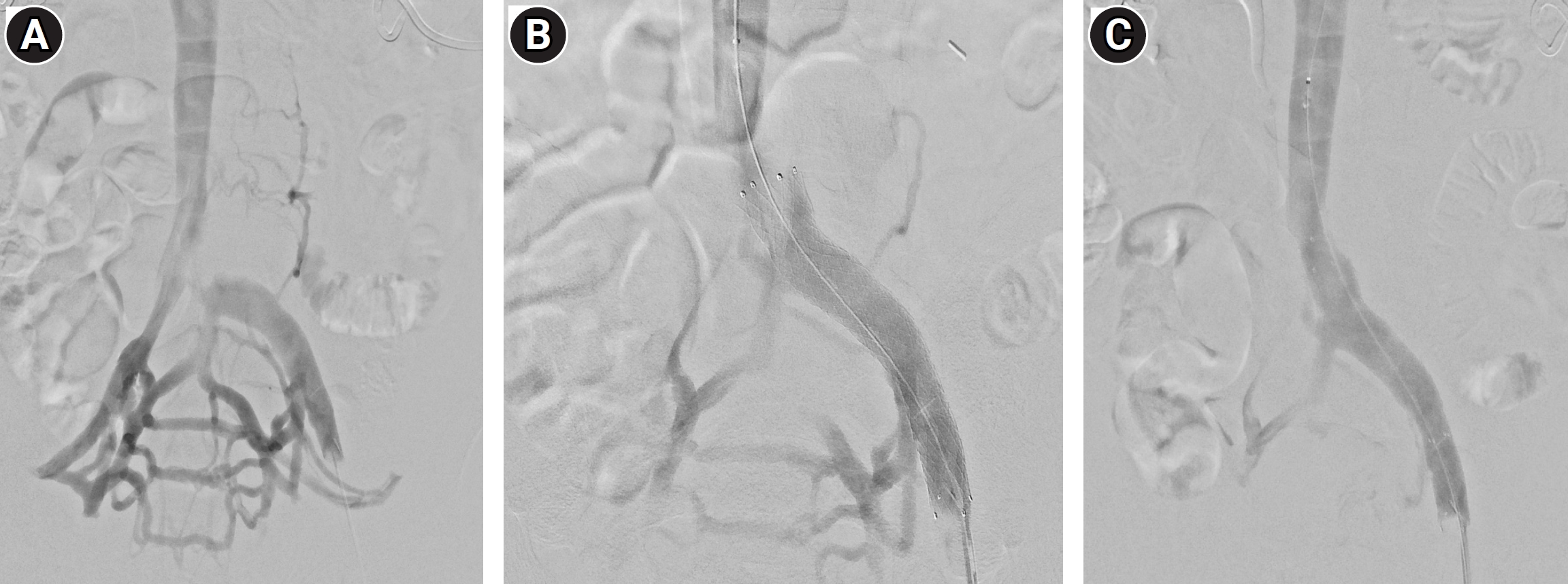
Fig. 5.Importance of adequate distal inflow in a 74-year-old man with a 10-day history of left leg swelling. (A) Initial venography demonstrates severe stenosis of the left common iliac vein with collateral venous drainage. (B) Iliac vein stenting followed by balloon angioplasty results in satisfactory expansion of the proximal stent. (C) Venography demonstrates additional chronic post-thrombotic stenosis involving the distal external iliac vein and common femoral vein, with persistent collateral venous filling. (D) A second stent is placed to provide complete coverage of the distal diseased segment, resulting in disappearance of the collateral veins and restoration of brisk venous outflow. An IVC filter was placed prophylactically because of concern for pulmonary thromboembolism during the procedure.
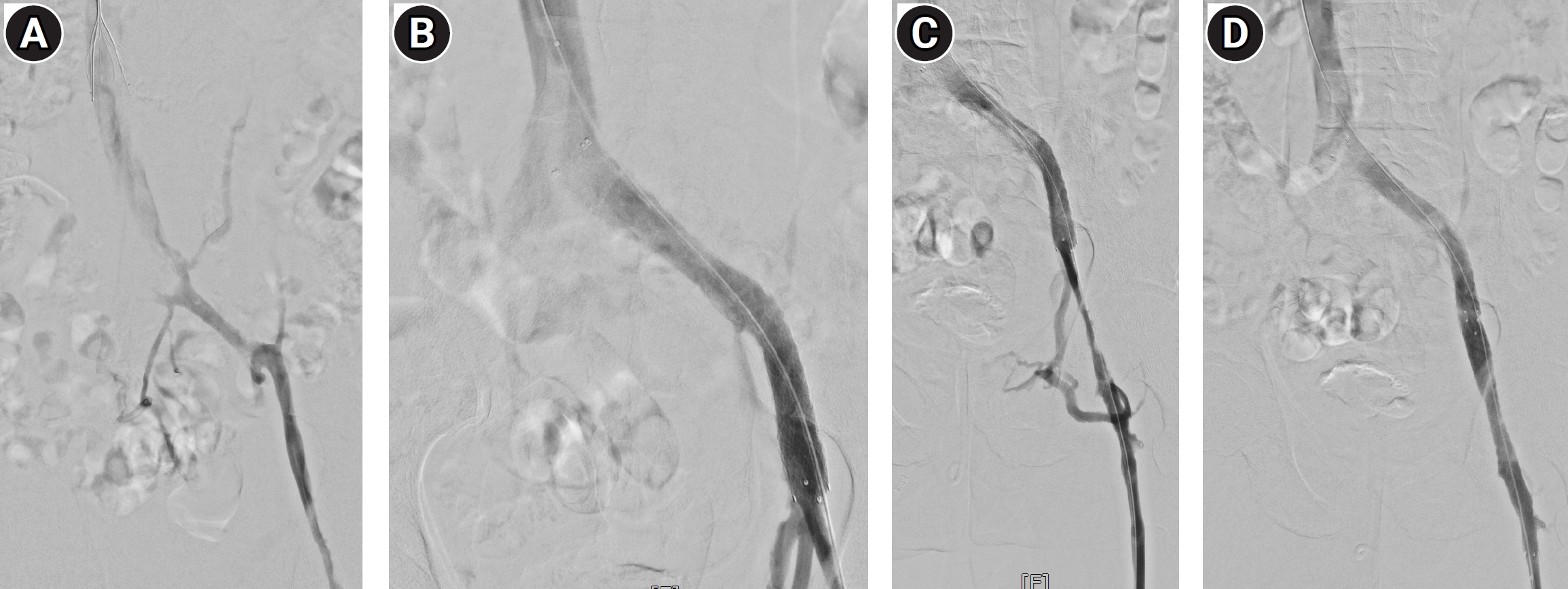
Table 1.Practical approach to iliac vein stenting according to clinical phenotype
Table 1.
|
Clinical phenotype |
When to consider intervention |
Key assessment |
Stenting strategy |
Practical point |
|
Uncomplicated NIVL |
Significant symptoms plausibly attributable to iliac outflow obstruction after competing causes are considered |
Duplex US; CT/MR venography; multiplanar venography; IVUS when available |
Cover the complete lesion; size to a normal reference vein; minimize unnecessary contralateral CIV ostial coverage |
Do not treat anatomic compression alone; clinical response is the primary goal |
|
Acute iliofemoral DVT |
Selected patients undergoing early thrombus removal with significant residual fixed iliac obstruction |
Thrombus clearance followed by venography/IVUS reassessment; assess inflow and residual thrombus |
Stent a persistent culprit iliac lesion after adequate thrombus clearance |
Anticoagulation follows VTE principles; IVC filter only in selected circumstances |
|
Chronic post-thrombotic obstruction |
Clinically significant PTS with imaging-confirmed iliofemoral obstruction |
Define full disease extent and inflow; assess CFV/femoral/profunda pathways |
Reconstruct the entire diseased segment; extend across the inguinal ligament when required to reach adequate inflow |
Patency is more dependent on inflow and thrombotic risk; antithrombotic therapy individualized |
Table 2.Major clinical studies relevant to iliac venous intervention
Table 2.
|
Study |
Design |
Population |
Key finding |
Practical implication |
|
VIDIO [19] |
Prospective multicenter diagnostic/clinical outcome study |
Patients undergoing iliofemoral venous stenting |
IVUS-derived stenosis predicted clinical improvement better than venography; area stenosis >54% and NIVL diameter stenosis >61% were associated with improvement |
Use IVUS measurements as supportive, not absolute, treatment thresholds |
|
ATTRACT [17] |
Randomized trial |
Acute proximal DVT; iliofemoral subgroup included |
Routine pharmacomechanical catheter-directed thrombolysis did not reduce overall PTS and increased bleeding |
Select patients for thrombus removal rather than treating all proximal DVT |
|
ABRE [33] |
Prospective multicenter single-arm study |
Symptomatic iliofemoral venous outflow obstruction |
Durable 3-year patency; outcomes differed by disease phenotype, with higher patency in NIVL than PTS |
Disease phenotype strongly influences expected patency |
|
Venovo [34] |
Prospective multicenter single-arm study |
Symptomatic iliac/femoral venous obstruction |
Sustained midterm patency and clinical improvement with a dedicated venous stent |
Supports dedicated venous stenting; avoid cross-trial device ranking |
|
VIRTUS [35] |
Prospective multicenter single-arm study |
Symptomatic iliofemoral venous obstruction |
Long-term results supported durability of a dedicated nitinol venous stent |
Interpret outcomes in light of lesion phenotype and study definitions |
|
VIVO [36,37] |
Prospective multicenter single-arm study |
Symptomatic iliofemoral venous outflow obstruction |
Favorable 1- and 3-year outcomes with the Zilver Vena venous stent |
Dedicated-stent evidence is supportive but not head-to-head |
|
C-TRACT [38] |
Randomized controlled trial |
Moderate/severe PTS with iliac-vein obstruction |
Endovascular therapy improved VCSS and venous disease-specific quality of life at 6 months but increased bleeding |
In PTS, balance symptomatic benefit against procedural and bleeding risk |
References
- 1. May R, Thurner J. The cause of the predominantly sinistral occurrence of thrombosis of the pelvic veins. Angiology. 1957;8:419-427. https://doi.org/10.1177/000331975700800505
- 2. Cockett FB, Thomas ML. The iliac compression syndrome. Br J Surg. 1965;52:816-821. https://doi.org/10.1002/bjs.1800521028
- 3. Desai KR, Sabri SS, Elias S, Gagne PJ, Garcia MJ, Gibson K, et al. Consensus statement on the management of nonthrombotic iliac vein lesions from the VIVA Foundation, the American Venous Forum, and the American Vein and Lymphatic Society. Circ Cardiovasc Interv. 2024;17:e014160. https://doi.org/10.1161/CIRCINTERVENTIONS.124.014160
- 4. Raju S, Neglen P. High prevalence of nonthrombotic iliac vein lesions in chronic venous disease: a permissive role in pathogenicity. J Vasc Surg. 2006;44:136-143. https://doi.org/10.1016/j.jvs.2006.02.065
- 5. Mousa AY, AbuRahma AF. May-Thurner syndrome: update and review. Ann Vasc Surg. 2013;27:984-995. https://doi.org/10.1016/j.avsg.2013.05.001
- 6. Kibbe MR, Ujiki M, Goodwin AL, Eskandari M, Yao J, Matsumura J. Iliac vein compression in an asymptomatic patient population. J Vasc Surg. 2004;39:937-943. https://doi.org/10.1016/j.jvs.2003.12.032
- 7. Oguzkurt L, Ozkan U, Ulusan S, Koc Z, Tercan F. Compression of the left common iliac vein in asymptomatic subjects and patients with left iliofemoral deep vein thrombosis. J Vasc Interv Radiol. 2008;19:366-370. https://doi.org/10.1016/j.jvir.2007.09.007
- 8. Marston W, Fish D, Unger J, Keagy B. Incidence of and risk factors for iliocaval venous obstruction in patients with active or healed venous leg ulcers. J Vasc Surg. 2011;53:1303-1308. https://doi.org/10.1016/j.jvs.2010.10.120
- 9. Raju S, Kirk OK, Jones TL. Endovenous management of venous leg ulcers. J Vasc Surg Venous Lymphat Disord. 2013;1:165-172. https://doi.org/10.1016/j.jvsv.2012.09.006
- 10. Vedantham S, Weinberg I, Desai KR, Winokur R, Kolli KP, Patel S, et al. Society of Interventional Radiology position statement on the management of chronic iliofemoral venous obstruction with endovascular placement of metallic stents. J Vasc Interv Radiol. 2023;34:1643-1657. https://doi.org/10.1016/j.jvir.2023.06.013
- 11. Mahnken AH, Thomson K, de Haan M, O'Sullivan GJ. CIRSE standards of practice guidelines on iliocaval stenting. Cardiovasc Intervent Radiol. 2014;37:889-897. https://doi.org/10.1007/s00270-014-0875-4
- 12. Neglen P, Hollis KC, Olivier J, Raju S. Stenting of the venous outflow in chronic venous disease: long-term stent-related outcome, clinical, and hemodynamic result. J Vasc Surg. 2007;46:979-990. https://doi.org/10.1016/j.jvs.2007.06.046
- 13. Hartung O, Loundou AD, Barthelemy P, Arnoux D, Boufi M, Alimi YS. Endovascular management of chronic disabling ilio-caval obstructive lesions: long-term results. Eur J Vasc Endovasc Surg. 2009;38:118-124. https://doi.org/10.1016/j.ejvs.2009.03.004
- 14. Chen F, Deng J, Hu XM, Zhou WM. Compression of the right iliac vein in asymptomatic subjects and patients with iliofemoral deep vein thrombosis. Phlebology. 2016;31:471-480. https://doi.org/10.1177/0268355515598700
- 15. Rossi FH, Kambara AM, Izukawa NM, Rodrigues TO, Rossi CB, Sousa AG, et al. Randomized double-blinded study comparing medical treatment versus iliac vein stenting in chronic venous disease. J Vasc Surg Venous Lymphat Disord. 2018;6:183-191. https://doi.org/10.1016/j.jvsv.2017.11.003
- 16. Razavi MK, Jaff MR, Miller LE. Safety and effectiveness of stent placement for iliofemoral venous outflow obstruction: systematic review and meta-analysis. Circ Cardiovasc Interv. 2015;8:e002772. https://doi.org/10.1161/CIRCINTERVENTIONS.115.002772
- 17. Vedantham S, Goldhaber SZ, Julian JA, Kahn SR, Jaff MR, Cohen DJ, et al. Pharmacomechanical catheter-directed thrombolysis for deep-vein thrombosis. N Engl J Med. 2017;377:2240-2252. https://doi.org/10.1056/NEJMoa1615066
- 18. Metzger PB, Rossi FH, Kambara AM, Izukawa NM, Saleh MH, Pinto IM, et al. Criteria for detecting significant chronic iliac venous obstructions with duplex ultrasound. J Vasc Surg Venous Lymphat Disord. 2016;4:18-27. https://doi.org/10.1016/j.jvsv.2015.07.002
- 19. Gagne PJ, Gasparis A, Black S, Thorpe P, Passman M, Vedantham S, et al. Analysis of threshold stenosis by multiplanar venogram and intravascular ultrasound examination for predicting clinical improvement after iliofemoral vein stenting in the VIDIO trial. J Vasc Surg Venous Lymphat Disord. 2018;6:48-56. https://doi.org/10.1016/j.jvsv.2017.07.009
- 20. Neglen P, Raju S. Intravascular ultrasound scan evaluation of the obstructed vein. J Vasc Surg. 2002;35:694-700. https://doi.org/10.1067/mva.2002.121127
- 21. Black SA, Gohel M, de Graaf R, Gagne P, Silver M, Fleck B, et al. Editor's choice - management of lower extremity venous outflow obstruction: results of an International Delphi Consensus. Eur J Vasc Endovasc Surg. 2024;67:341-350. https://doi.org/10.1016/j.ejvs.2023.09.044
- 22. Neglen P, Raju S. Balloon dilation and stenting of chronic iliac vein obstruction: technical aspects and early clinical outcome. J Endovasc Ther. 2000;7:79-91. https://doi.org/10.1177/152660280000700201
- 23. Li N, Mendoza F, Rugonyi S, Farsad K, Kaufman JA, Jahangiri Y, et al. Venous biomechanics of angioplasty and stent placement: implications of the Poisson effect. J Vasc Interv Radiol. 2020;31:1348-1356. https://doi.org/10.1016/j.jvir.2020.02.033
- 24. Murphy EH, Johns B, Varney E, Buck W, Jayaraj A, Raju S. Deep venous thrombosis associated with caval extension of iliac stents. J Vasc Surg Venous Lymphat Disord. 2017;5:8-17. https://doi.org/10.1016/j.jvsv.2016.09.002
- 25. Caliste XA, Clark AL, Doyle AJ, Cullen JP, Gillespie DL. The incidence of contralateral iliac venous thrombosis after stenting across the iliocaval confluence in patients with acute or chronic venous outflow obstruction. J Vasc Surg Venous Lymphat Disord. 2014;2:253-259. https://doi.org/10.1016/j.jvsv.2013.12.007
- 26. Le TB, Lee TK, Park KM, Jeon YS, Hong KC, Cho SG. Contralateral deep vein thrombosis after iliac vein stent placement in patients with May-Thurner syndrome. J Vasc Interv Radiol. 2018;29:774-780. https://doi.org/10.1016/j.jvir.2018.01.771
- 27. Duarte-Gamas L, Rocha-Neves JP, Pereira-Neves A, Dias-Neto M, Baekgaard N. Contralateral deep vein thrombosis after stenting across the iliocaval confluence in chronic venous disease: a systematic review. Phlebology. 2020;35:221-230. https://doi.org/10.1177/0268355519889873
- 28. Jacob A, Theis C, Betancourt A, Calderon D, Marston WA. Postinsertion iliac vein stent position and relationship to contralateral deep vein thrombosis after unilateral iliac vein stenting. J Vasc Surg Venous Lymphat Disord. 2026;14:102577. https://doi.org/10.1016/j.jvsv.2026.102577
- 29. Machado H, Sousa J, Mansilha A. The impact of venous stenting across the inguinal ligament on primary patency: a systematic review. Int Angiol. 2021;40:270-276. https://doi.org/10.23736/S0392-9590.21.04663-0
- 30. Ye K, Shi H, Yin M, Qin J, Yang X, Liu X, et al. Treatment of femoral vein obstruction concomitant with iliofemoral stenting in patients with severe post-thrombotic syndrome. Eur J Vasc Endovasc Surg. 2018;55:222-228. https://doi.org/10.1016/j.ejvs.2017.11.029
- 31. Raju S, Luke C, Powell T, Saleem T, Jayaraj A. Extension of iliac vein stent into the profunda femoral vein for salvage. J Vasc Surg Venous Lymphat Disord. 2022;10:1059-1065. https://doi.org/10.1016/j.jvsv.2022.03.016
- 32. Friedrich de Wolf MA, Arnoldussen CW, Grommes J, Hsien SG, Nelemans PJ, de Haan MW, et al. Minimally invasive treatment of chronic iliofemoral venous occlusive disease. J Vasc Surg Venous Lymphat Disord. 2013;1:146-153. https://doi.org/10.1016/j.jvsv.2012.07.002
- 33. Black S, Sapoval M, Dexter DJ, Gibson K, Kolluri R, Razavi M, et al. Three-year outcomes of the ABRE venous self-expanding stent system in patients with symptomatic iliofemoral venous outflow obstruction. J Vasc Interv Radiol. 2024;35:664-675. https://doi.org/10.1016/j.jvir.2024.01.030
- 34. Dake MD, O'Sullivan G, Shammas NW, Lichtenberg M, Mwipatayi BP, Settlage RA, et al. Three-year results from the Venovo venous stent study for the treatment of iliac and femoral vein obstruction. Cardiovasc Intervent Radiol. 2021;44:1918-1929. https://doi.org/10.1007/s00270-021-02975-2
- 35. Razavi MK, Gagne P, Black S, Sadek M, Nicolini P, Weinberg I, et al. Midterm and long-term outcomes following dedicated endovenous nitinol stent placement for symptomatic iliofemoral venous obstruction: three- to 5-year results of the VIRTUS study. J Vasc Interv Radiol. 2022;33:1485-1491. https://doi.org/10.1016/j.jvir.2022.08.028
- 36. Hofmann LR, Gagne P, Brown JA, Saunders A, Comerota A; VIVO Study Investigators. Twelve-month end point results from the evaluation of the Zilver Vena venous stent in the treatment of symptomatic iliofemoral venous outflow obstruction (VIVO clinical study). J Vasc Surg Venous Lymphat Disord. 2023;11:532-541. https://doi.org/10.1016/j.jvsv.2022.12.066
- 37. Comerota AJ, Gagne P, Brown JA, Segbefia E, Hofmann LV; VIVO Study Investigators. Final 3-year study outcomes from the evaluation of the Zilver Vena venous stent for the treatment of symptomatic iliofemoral venous outflow obstruction (The VIVO Clinical Study). J Vasc Interv Radiol. 2024;35:834-845. https://doi.org/10.1016/j.jvir.2024.02.025
- 38. Vedantham S, Kahn SR, Marston WA, Weinberg I, Sista AK, Magnuson EA, et al. Endovascular therapy for post-thrombotic syndrome: a randomized trial. N Engl J Med. 2026;394:2293-2304. https://doi.org/10.1056/NEJMoa2519001
- 39. Milinis K, Thapar A, Shalhoub J, Davies AH. Antithrombotic therapy following venous stenting: international Delphi consensus. Eur J Vasc Endovasc Surg. 2018;55:537-544. https://doi.org/10.1016/j.ejvs.2018.01.007
- 40. Notten P, Ten Cate H, Ten Cate-Hoek AJ. Postinterventional antithrombotic management after venous stenting of the iliofemoral tract in acute and chronic thrombosis: a systematic review. J Thromb Haemost. 2021;19:753-796. https://doi.org/10.1111/jth.15197