Abstract
Extrahepatic collateral supply from the Sappey superior artery of the right internal thoracic artery (RITA) is an uncommon but important source of perfusion to HCC located in segment IV. Preprocedural CT demonstrated a large segment IV HCC supplied by the Sappey superior artery. Initial transfemoral angiography confirmed tumor vascularization but failed to achieve superselective catheterization because of marked tortuosity of the aortic arch. The procedure was converted to a right transradial approach, which provided stable access to the RITA and enabled successful superselection of the Sappey superior artery. Y-90 radioembolization via the Sappey superior artery and hepatic artery was completed without complications.
-
Keywords: Carcinoma, hepatocellular; Radioembolization, therapeutic; Mammary arteries; Radial artery; Yttrium radioisotopes
Introduction
Extrahepatic collateral arterial supply is frequently encountered in HCC, particularly in large or subcapsular tumors [
1]. Among these collateral pathways, the inferior phrenic artery is the most common source [
1]; however, the internal thoracic artery and its branches, including the Sappey superior artery, may also contribute to tumor perfusion, especially for lesions located in segment IV or abutting the anterior hepatic surface [
2,
3]. Recognition of these vessels is essential because incomplete treatment of collateral supply may compromise the efficacy of locoregional therapy.
Yttrium-90 (Y-90) transarterial radioembolization has increasingly been performed through parasitized extrahepatic arteries when superselective catheterization can be safely achieved. Recent reports have demonstrated the feasibility of radioembolization via the inferior phrenic artery, internal thoracic artery, and adrenal artery [
4-
9].
We report a case of successful Y-90 radioembolization performed through the Sappey superior artery originating from the right internal thoracic artery (RITA). Because severe tortuosity of the aortic arch prevented superselective catheterization from a transfemoral approach, conversion to a right transradial approach enabled stable catheterization and successful treatment.
Case Report
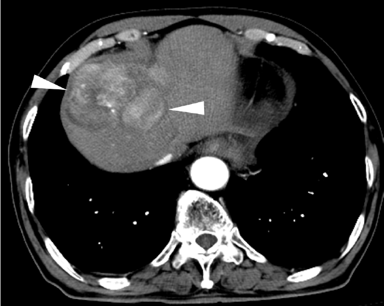
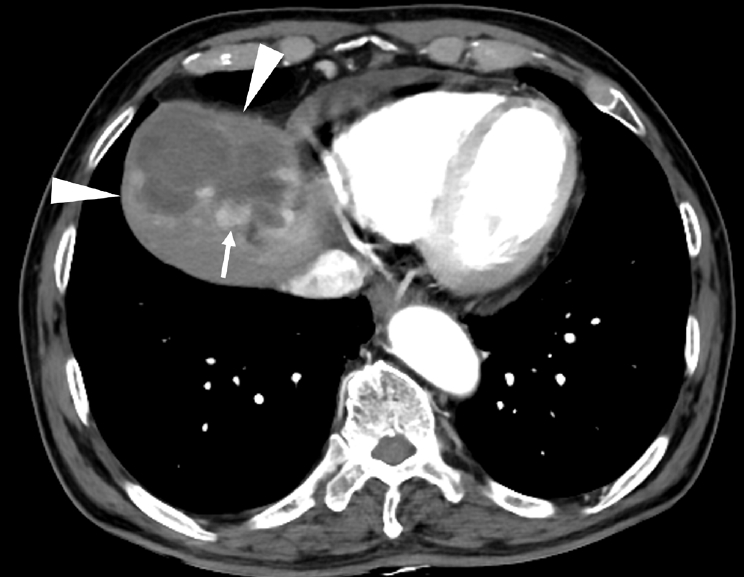
Institutional Review Board approval was waived for this case report in accordance with the policy of Seoul National University Hospital. An 86-year-old man presented with an 8.5-cm-sized single nodular HCC located in hepatic segment IV (
Fig. 1). He had preserved liver function with Child-Pugh class A5. Preprocedural laboratory tests showed total bilirubin, 0.8 mg/dL; albumin, 4.6 g/dL; prothrombin time/INR, 1.03; platelet count, 244 × 10³/µL; serum creatinine, 0.7 mg/dL; serum α-fetoprotein, 5.6 ng/mL; and protein induced by vitamin K antagonist-II, 305 mAU/mL. Contrast-enhanced CT demonstrated a parasitized extrahepatic arterial supply arising from the Sappey superior artery, which originated from the RITA and coursed through the falciform ligament to supply the tumor (
Video 1). Because of the absence of dysmorphic intratumoral vessel, same-day radioembolization with resin microspheres (SIR-Spheres, SIRTEX, Woburn, MA, USA) was planned.
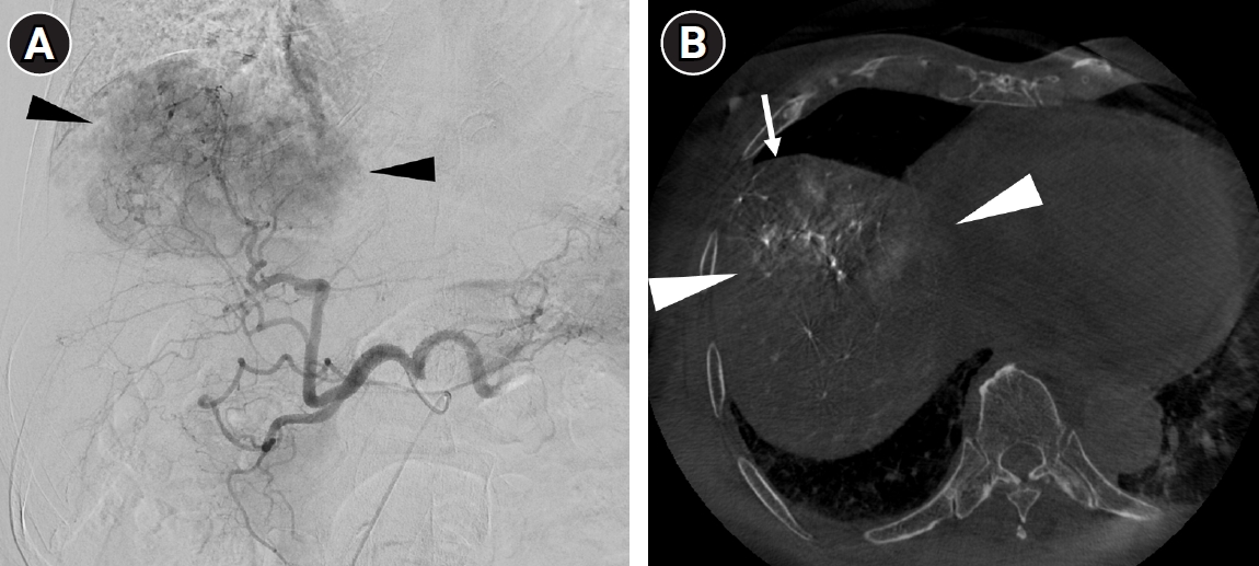
The procedure was initially performed via a right common femoral artery approach with a 5-Fr catheter (RH, Cook, Bloomington, IN, USA). Celiac angiography and cone-beam CT obtained at the proper hepatic artery revealed a hypervascular tumor in segment IV (
Fig. 2A), but focal unenhancement of the anterior portion of the tumor was noted on cone-beam CT (
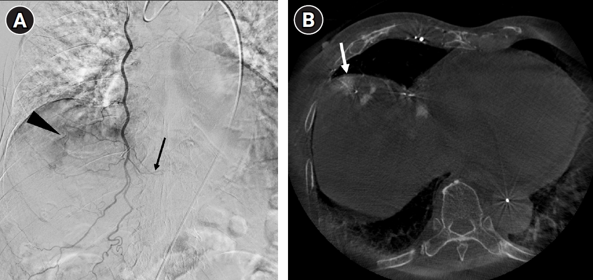
Fig. 2B), which suggested the presence of extrahepatic collateral blood supply. Selective angiography and cone-beam CT of the RITA demonstrated that the anterior portion of the tumor was supplied by the Sappey superior artery of the RITA (
Fig. 3).
99mTc-macroaggregated albumin (MAA) was injected via the left hepatic artery, but not via the RITA. The patient was transferred to the department of nuclear medicine to obtain SPECT/CT, resulting in a lung shunt fraction of 3.17% and a tumor-to-normal ratio of 3.5. The total liver volume was 1,041 mL, of which 419 mL was planned for treatment. The total tumor volume was 181 mL, with 168 mL supplied by the segment IV artery and 13 mL by the RITA, as determined by cone-beam CT.
The virtual tumor absorbed dose (vTAD) was calculated under the assumption that all radioactive microspheres were deposited within the tumor. Based on the tumor volume of 13 mL supplied by the RITA, an activity of 0.19 GBq was required to achieve the target vTAD of 700 Gy. Therefore, the prescribed activity for the RITA was set at 0.2 GBq. For treatment via the segment IV artery, partition dosimetry was used. Given a treated liver volume of 406 mL, a treated tumor volume of 168 mL, and a tumor-to-normal ratio of 3.5, an activity of 2.45 GBq was required to achieve the target tumor absorbed dose (TAD) of 500 Gy. Accordingly, the prescribed activity for the segment IV artery was set at 2.5 GBq.
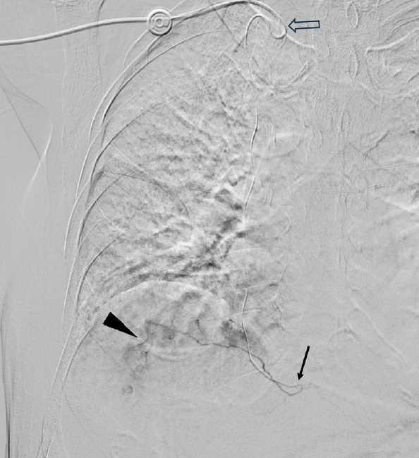
The patient was sent back to the angiography suite, and then the treatment procedure was initiated 30 minutes after SPECT/CT. Treatment via the Sappey superior artery was first attempted. However, because of marked elongation and tortuosity of the aortic arch, advancement of the microcatheter into the Sappey superior artery was technically unsuccessful. To overcome this limitation, vascular access via the right radial artery was added using a 5-Fr sheath (Prelude, Merit Medical, South Jordan, UT, USA). Through the transradial route, the RITA was readily catheterized with a 5-Fr Omni catheter (A&A, Seoul, Korea), which improved coaxial support and allowed advancement of a 1.7-Fr microcatheter (Progreat Lambda, Terumo, Tokyo, Japan) into the Sappey superior artery (
Fig. 4). Resin microspheres (SIR-Spheres, SIRTEX) of 0.2 GBq were delivered via the Sappey superior artery. Through the femoral approach, the segment IV artery was treated with resin microspheres of 2.5 GBq. Residual activity was measured after the procedure, and the actual delivered radiation activity was 0.18 GBq and 2.46 GBq via the Sappey superior artery and segment IV artery, respectively. Post-treatment PET/CT imaging was obtained the next morning, which demonstrated hot radiation activity within the entire tumor and no significant extrahepatic uptake, and mean TAD was calculated as 586 Gy.
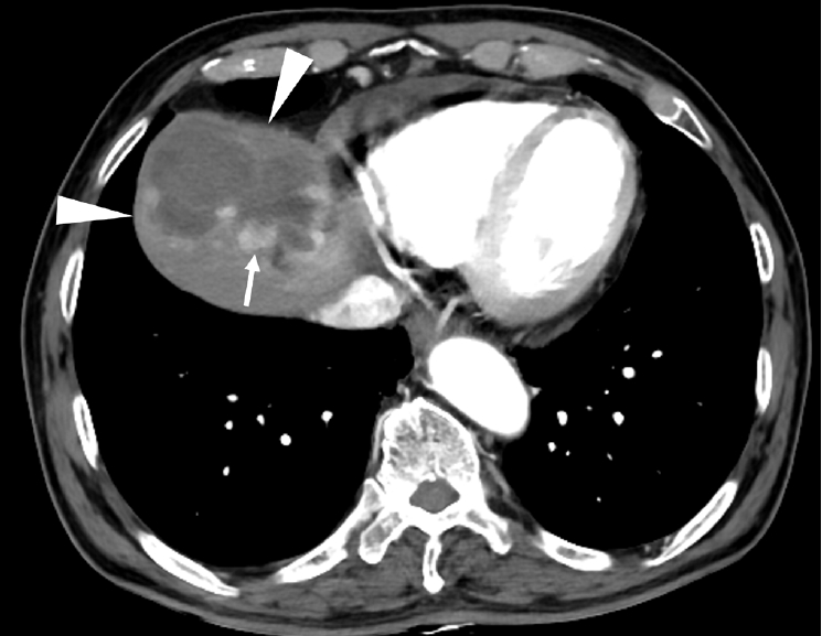
The patient tolerated the procedure well without access-site complications or clinically significant adverse events. Follow-up CT scan 6 months after radioembolization demonstrated partial response with minimal residual tumor by modified Response Evaluation Criteria in Solid Tumors (
Fig. 5). CT scan 8 months after radioembolization demonstrated a slight increase in the residual viable tumor, and a second radioembolization was performed. During the second procedure, selective angiography and cone-beam CT showed that the residual viable tumor was supplied exclusively by the segment IV artery, with no tumor supplied by the RITA. A total activity of 1.1 GBq was administered through the segment IV artery. Post-treatment PET/CT demonstrated a TAD of 578 Gy. Until the last follow-up, no complications occurred.
Discussion
The Sappey superior artery is an important but underrecognized collateral pathway supplying HCCs located in the anterior superior liver, particularly segment IV lesions adjacent to the falciform ligament [
2]. Previous anatomic studies have shown that this vessel may arise from the internal thoracic artery and communicate with the inferior phrenic circulation through the diaphragm [
2]. Consequently, careful review of preprocedural cross-sectional imaging and angiography is required to identify this collateral source [
10].
Although several reports have described transarterial chemoembolization or radioembolization through parasitized internal thoracic or extrahepatic arteries, experience with Y-90 delivery through the Sappey superior artery remains extremely limited [
8]. Complete treatment of all arterial supplies is particularly important for radioembolization when curative or ablative intent is pursued, because omission of a collateral feeder may leave viable tumor tissue untreated.
The RITA gives rise to the anterior intercostal arteries and the superior epigastric artery, which supply the anterior chest/abdominal wall and overlying skin. Consequently, non-selective radioembolization through the RITA carries a substantial risk of radiation dermatitis. In contrast, the Sappey superior artery supplies the tumor and the diaphragm without providing arterial supply to the skin. Therefore, superselective radioembolization through the Sappey superior artery is unlikely to result in radiation dermatitis. Because resin microspheres have a relatively high particle load, microsphere reflux may occur during administration. If reflux occurs into the non-target branches of the RITA, radiation dermatitis may develop. To minimize this risk, the author continuously infused a 1:1 mixture of contrast medium and 5% dextrose through the injection line at a very slow rate, allowing real-time monitoring of antegrade flow and immediate detection of any reflux. This technique facilitated maintenance of antegrade flow throughout the procedure and helped prevent microsphere reflux into non-target vessels.
This case also highlights an important technical consideration regarding vascular access. Transfemoral catheterization of the RITA may be difficult in elderly patients with severe elongation and tortuosity of the aortic arch. In contrast, a right transradial approach provides a shorter and more favorable trajectory to the RITA, improving catheter stability and facilitating distal microcatheter advancement. In this patient, conversion from femoral to radial access transformed an unsuccessful procedure into successful superselective catheterization and definitive treatment.
The present case supports two practical messages. First, the Sappey superior artery should be actively sought in patients with segment IV HCC demonstrating incomplete hepatic arterial perfusion. Second, when transfemoral access fails because of unfavorable aortic anatomy, a transradial approach should be considered as an effective alternative for selective catheterization of the RITA and its distal branches.
Appropriate patient selection is essential for safe implementation of same-day radioembolization. Based on previous studies and our prospective ISTAR-01 protocol [
11,
12], imaging findings suggestive of elevated lung shunting include hepatic vein invasion, early hepatic vein enhancement on arterial-phase CT or MRI, the presence of transjugular intrahepatic portosystemic shunt, and dysmorphic intratumoral vessels measuring ≥3 mm. Marked portal vein enhancement on arterial-phase CT or MRI indicates a massive arterioportal shunt, which is generally considered a contraindication to radioembolization because of the risk of unintended whole-liver irradiation. Therefore, the ideal candidates for same-day radioembolization are patients without any imaging features suggestive of high lung shunting and without evidence of a massive arterioportal shunt on preprocedural imaging. Even in patients without these imaging findings, same-day treatment should proceed only after MAA-based dosimetry confirms that an adequate TAD can be delivered while maintaining the estimated lung dose within accepted safety limits.
In conclusion, Y-90 radioembolization through the Sappey superior artery arising from the RITA is feasible and can be safely performed when meticulous superselective catheterization is achieved. In patients with severe aortic arch tortuosity, the right transradial approach may provide superior catheter stability compared with the transfemoral approach and facilitate successful treatment of HCC supplied by this uncommon extrahepatic collateral artery.
Conflict of interest
No potential conflict of interest relevant to this article was reported.
Funding
None.
Acknowledgments
None.
Author contributions
The author conducted all aspects of the study.
Data availability statement
The datasets generated or analyzed during the study are available from the corresponding author on reasonable request.
Supplementary material
Video 1.
The superior artery (arrowhead) can be traced from the right internal thoracic artery to the tumor on a serial image of a CT scan.
Fig. 1.CT scan shows a large enhancing tumor (arrowheads) in liver segment IV.
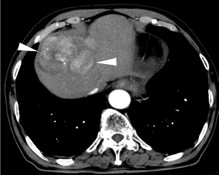
Fig. 2.(A) Celiac angiogram shows large tumor blush (arrowheads) supplied by the segment IV artery. (B) Axial image of cone-beam CT obtained at the proper hepatic artery shows tumor blush (arrowheads) in the liver dome. The anterior part (arrow) of the tumor was not enhanced.
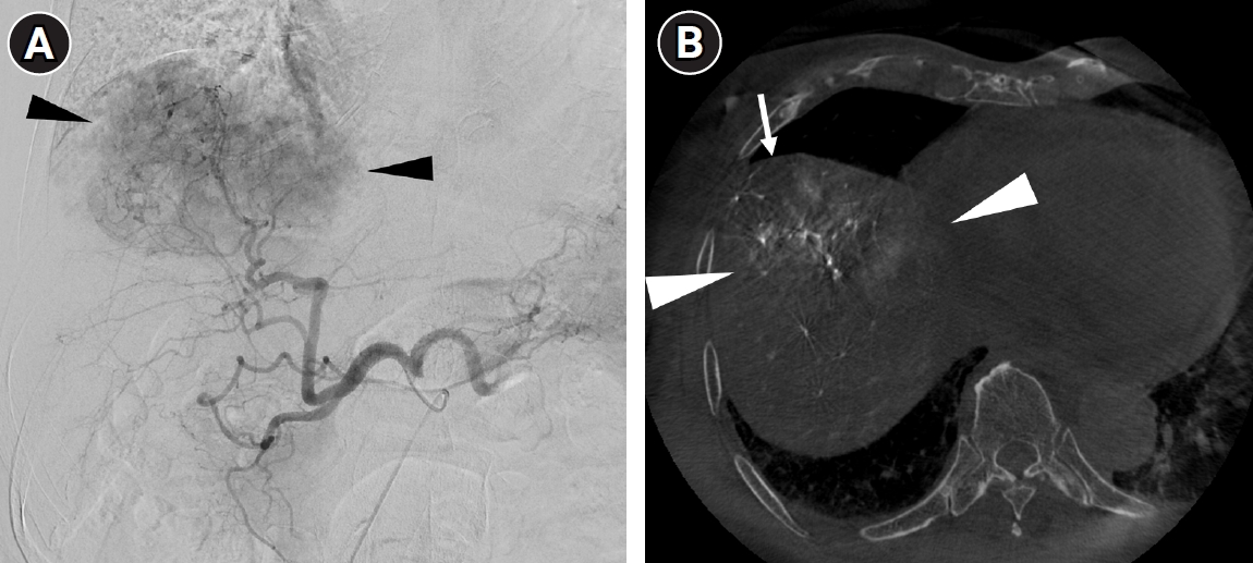
Fig. 3.(A) Right internal thoracic angiogram shows tumor blush (arrowhead) supplied by Sappey superior artery (arrow) from the right internal thoracic artery. (B) Axial image of cone-beam CT obtained at the right internal thoracic artery shows tumor blush (arrow) in the anterior part of the tumor.
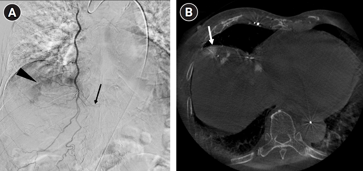
Fig. 4.Selective angiogram of the Sappey superior artery shows tumor blush (arrowhead). Note a microcatheter tip (arrow) in the Sappey superior artery and a 5-Fr catheter (open arrow) inserted via the right radial artery.
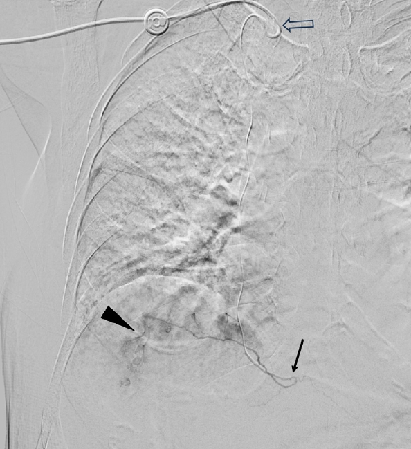
Fig. 5.CT scan 6 months after radioembolization shows small residual enhancing foci (arrow) within the treated tumor (arrowheads).
References
- 1. Kim HC, Chung JW, Lee W, Jae HJ, Park JH. Recognizing extrahepatic collateral vessels that supply hepatocellular carcinoma to avoid complications of transcatheter arterial chemoembolization. Radiographics. 2005;25 Suppl 1:S25-S39. https://doi.org/10.1148/rg.25si055508
- 2. Kim HC, Miyayama S, Choi JW, Kim GM, Chung JW. Hepatocellular carcinoma supplied by the inferior phrenic artery or cystic artery: anatomic and technical considerations. Radiographics. 2023;43:e220076. https://doi.org/10.1148/rg.220076
- 3. Kim HC, Chung JW, Choi SH, Jae HJ, Lee W, Park JH. Internal mammary arteries supplying hepatocellular carcinoma: vascular anatomy at digital subtraction angiography in 97 patients. Radiology. 2007;242:925-932. https://doi.org/10.1148/radiol.2423060220
- 4. Kim HC, Kim YJ, Paeng JC, Chung JW. Yttrium-90 radioembolization of the right inferior phrenic artery in 20 patients with hepatocellular carcinoma. J Vasc Interv Radiol. 2018;29:556-563. https://doi.org/10.1016/j.jvir.2017.10.010
- 5. Kim HC, Kim DH, Suh M, Paeng JC, Choi JW. High-dose radioembolization limited by lung shunt for hepatocellular carcinoma supplied by the inferior phrenic artery. J Vasc Interv Radiol. 2025;36:1427-1435. https://doi.org/10.1016/j.jvir.2025.05.027
- 6. Vidal LL, Frey GT, Ritchie C, Lewis A, Paz-Fumagalli R, McKinney M, et al. Ablative transarterial radioembolization of a parasitized adrenal artery for the treatment of hepatocellular carcinoma. J Vasc Interv Radiol. 2019;30:473-476. https://doi.org/10.1016/j.jvir.2018.10.022
- 7. Arotzarena G, Toskich BB, Lewis AR, Paz-Fumagalli R. Ablative radioembolization of hepatocellular carcinoma with total arterial supply originating from the superior adrenal artery achieved complete pathologic necrosis. Radiol Case Rep. 2023;18:196-199. https://doi.org/10.1016/j.radcr.2022.10.025
- 8. Alhussaini A, Qazi SA, AlZahrani YA. Successful transcatheter arterial radioembolization of hepatocellular carcinoma via left internal mammary artery: a case report. Cureus. 2022;14:e22954. https://doi.org/10.7759/cureus.22954
- 9. Okinedo I, Frageau J, Krishnarao A, Figueira T, Resnick N, Li NP. Yttrium-90 transarterial radioembolization of a hepatocellular carcinoma via parasitized adrenal and renal capsular arteries. J Vasc Interv Radiol. 2026;37:108739. https://doi.org/10.1016/j.jvir.2026.108739
- 10. Kim HC, Chung JW, Jae HJ, Jeon UB, Son KR, Park JH. Hepatocellular carcinoma: prediction of blood supply from an internal mammary artery with multi-detector row CT. J Vasc Interv Radiol. 2008;19:1419-1425. https://doi.org/10.1016/j.jvir.2008.06.015
- 11. Kim HC, Suh M, Paeng JC, Choi JW. Same-day versus multiday planning/treatment radioembolization with yttrium-90 resin microspheres in patients with liver cancer ≥5 cm. J Vasc Interv Radiol. 2025;36:2010-2020. https://doi.org/10.1016/j.jvir.2025.08.028
- 12. Kim HC, Lee IJ, Hyun DH, Kim GM. Same-day radioembolization for large hepatocellular carcinoma (≥5 cm) using yttrium-90 resin microspheres: protocol for a multicenter prospective registry study (ISTAR-01). Cardiovasc Intervent Radiol. (in press).