중심단어
EVAR, AngioJet, kissing stent technique, limb graft occlusion
한글 초록
복부대동맥류의 가장 잘 알려진 치료방법은 혈관내 치료이다. 하지만 복부대동맥이 꺽인 정도에 따라 이후에 인조혈관의 주체(主體)와분지부위에 꺽임이 생겨 원위부의 혈류가 느려지게 되고 그에 따라 혈전성 폐색이 생기는 원인이 되기도 한다. 이에 본 저자들은 혈관내 치료후 혈전성 폐색이 발생한 경험을 보고하고, 추가적인 시술로 극복해 낸 기술적 방법을 공유하고자 한다.
영문 초록
EVAR has been an established method for treatment of abdominal aortic aneurysms. However, the angulation of native abdominal aorta induce kinking at graft main body or limb graft, resulting thrombo-occlusion of distal branches. Herein, authors report our experience of thrombo-cclusion in iliac limb graft after EVAR, and share the overcoming method.
Introduction
복부대동맥류의 혈관 내 치료 후 발생한 장골 분지 인조혈관의 혈전성 폐색 치료에 대해 알아보고자 한다.
Case report
증례
78세/남자
임상소견
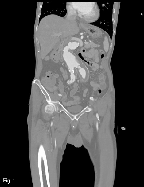
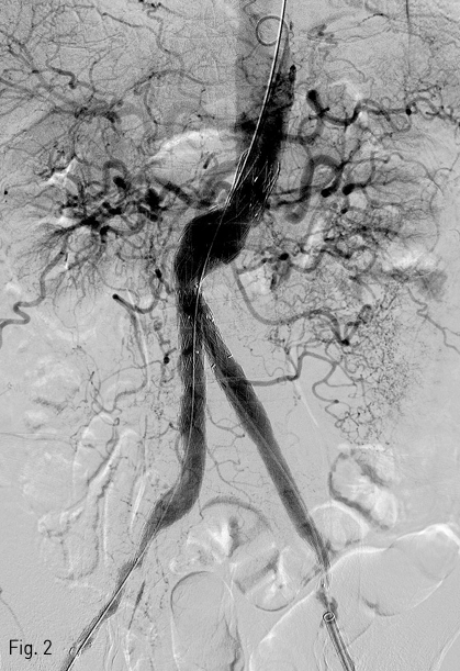
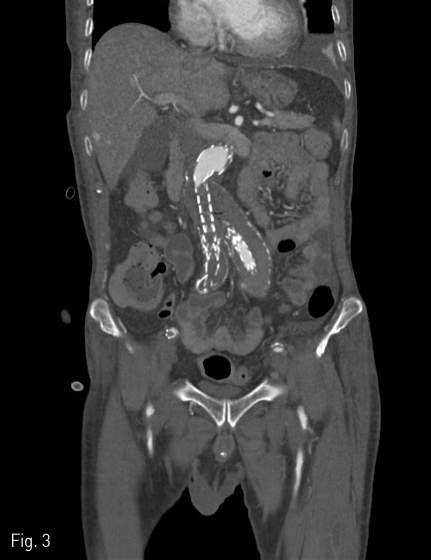
2018년 좌측발의 통증과 색깔변화를 주소로 내원하여 시행한 조영증강 CT(Fig. 1)상에서 발견된 복부대동맥 및 양측 장골 동맥류에 대해 InCraft(Cordis, Milpats, US)를 이용한 EVAR 치료(Fig. 2)를 받은 환자로, 술후 퇴원전 조영증강 CT(Fig. 3)상에서 우측 장골 분지 인조혈관이 폐색된 소견을 보였다.
진단명
Thrombocclusion in right iliac limb graft
시술방법 및 재료
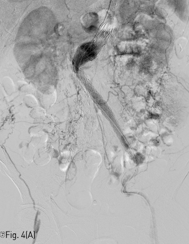
시술전 CT를 면밀히 검토하여 혈전성 폐색부위는 AngioJet (Boston scientific, Boston, US) 기구를 이용하여 개통시킨 다음, 인조혈관의 주체(main body) 부위와 분지 인조혈관사이의 이행대에 생긴 인조혈관 꺽임은 풍선확장식 스텐트를 이용하여 kissing balloon 기술로 설치하기로 계획하였다. 초음파 유도 하에 양측 총대퇴동맥을 천자하고 좌측 경로를 이용하여 5Fr pigtail catheter (COOK medical, Søborg, Denmark) 를 이용하여 혈관조영술을 시행하였다(Fig. 4A).
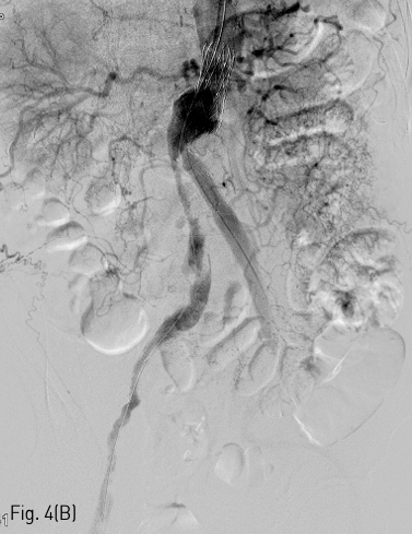
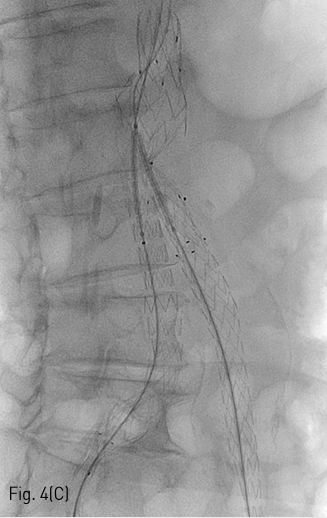
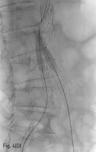
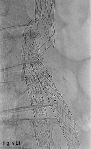
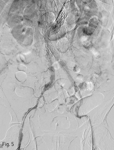
이후 우측 경로를 이용하여 AngioJet 기구를 진입시켜 2-3회에 걸쳐 혈전용해술을 시행하였고 우측 장골 분지의 내경은 회복되었다 (Fig. 4B). 이후 양측 경로에 들어있는 유도철사 (Terumo, Tokyo, Japan) 를 5Fr Berenstein catheter (Cordis, Milpitas, US) 를이용하여 Lunderquist (COOK medical, Søborg, Denmark) 유도철사로 교환한 뒤, Express LD premounted (Boston scientific, Boston, US) 스텐트를 우측은 8x37mm, 좌측은 10x37mm로 선택하여 꺽인 부위에 진입시켰다(Fig. 4C). 양측에서 조심스럽게 실시간으로 관찰하며 균형을 맞추어 스텐트를 설치하였다 (Fig. 4D). Mustang (Boston scientific, Boston, US) 9x60mm 크기의 풍선을 이용하여 설치 후 풍선확장술을 시행하여 최종적으로 스텐트의 모양을 완성하였다(Fig. 4E). 시술 직후 시행한 최종 혈관조영술 상에서 양측 분지로의 혈류가 정상적으로 회복되었으며, 혈류 속도도 양측이 균등함을 확인하였다(Fig. 5).
추적관찰
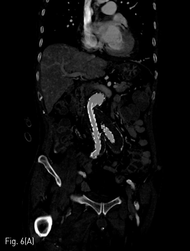
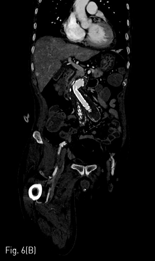
시술 1개월 뒤 시행한 조영증상 CT에서 양측 장골 분지 인조혈관 내강은 잘 유지되었다(Fig. 6A&B).
Fig 1
Pre EVAR CT angiography demonstrates abdominal aortic aneurysm with hostile neck.
Fig 2
Post EVAR angiography reveals successful exclusion of aortic aneurysm.
Fig 3
CT angiography obtained 4 days after EVAR shows thrombo-occlusive change of right limb graft.
Fig 4A
(A) Abdominal aortography shows well correlated findings with CT angiography.
Fig 4B
(B) Abdominal aortography demonstrates successful re-canalization of right limb graft after thrombolysis treatment with AngioJet device.
Fig 4C
(C,D) Spot radiography shows introducing stent by kissing method.
Fig 4D
(C,D) Spot radiography shows introducing stent by kissing method.
Fig 4E
(E) Single radiography demonstrates final result of kissing stent extension.
Fig 5
Abdominal aortography taken just after kissing stent insertion shows re-canalized right limb graft and equal flow via bilateral stent extension.
Fig 6A
(A,B) CT angiography obtained 1 month later the procedure shows patent intraluminal space of bilateral iliac limb graft.
Fig 6B
(A,B) CT angiography obtained 1 month later the procedure shows patent intraluminal space of bilateral iliac limb graft.
고찰
복부 대동맥류 뿐 아닌 다양한 동맥류에서 혈관내 치료 후 생기는 혈전성 폐색은 약 7.2%(6~28%)정도에서 나타나는 것으로 알려져 있으며, 복부 대동맥류 치료 후 생기는 부작용으로 흔히 알려진 endoleak보다 빈도(10%)는 적지만 드물지는 않다고 할 수 있다. 혈전성 폐색의 원인과 예측 요인은 다양한데, 이중 가장 중요한 요인은 대동맥류 목의 각도이다. 이 값이 60을 넘어가면 혈전성 폐색이 발생할 가능성이 그렇지 않은 군에 비해 2.3배 높아지며 재시술을 할 가능성도 38%정도 높아진다.
본 케이스의 환자 또한 대동맥류 목의 각도가 90도 이상이었으며, 비교적 이른 시일에 시행한 추적검사를 통해 빨리 발견하고 적절한 중재시술을 통해 성공적으로 치료할 수 있었다. 또한 스텐트 설치에 있어서, 두 스텐트가 서로 혈류 경쟁을 하기에 radial force의 평형을 잘 이룰 수 있는 kissing 스텐트 설치 기술이 필요하며 자가 확장식 스텐트보다는 풍선 확장식 스텐트를 선택하는 편이 용이하다.
인조혈관 제품의 특성과 환자의 해부학적 구조를 잘 파악하여 미리 발생할수 있는 혈전성 폐색에 대해 중재 시술 전에 진료과와 면밀한토의를 통해 의논하는 것이 최상의 결과를 도출해낼 수 있는 방법이라고 생각된다.
참고문헌
1. 대한인터벤션영상의학회. 인터벤션 영상의학 제2판. 일조각 2014:333-343
2. Cochennec F, Becquemin JP, Desgranges P, Allaire E, Kobeiter H, Roudot-Thoraval F. Limb Graft Occlusion Following EVAR: Clinical Pattern, Outcomes and Predictive Factors of Occurrence. Eur J Vasc Endovasc Surg 2007;34:59-65
3. Jeon YS, Cho YK, Song MG, et al..Clinical Outcomes of Endovascular Aneurysm Repair with the Kilt Technique for Abdominal Aortic Aneurysms with Hostile Aneurysm Neck Anatomy: A Korean Multicenter Retrospective Study. Cardiovasc Intervent Radiol 2018;41:554-563
4. Taudorf M, Jensen LP, Vogt KC, Gronvall J, Schroeder TV, L?nn L. Endograft Limb Occlusion in EVAR: Iliac Tortuosity Quantified by Three Different Indices on the Basis of Preoperative CTA. Eur J Vasc Endovasc Surg 2014;48:527-533
Citations
Citations to this article as recorded by