중심단어
Portosystemic shunt, hepatic encephalopathy, vein of Retzius, mesocaval shunt
한글 초록
알코올성 간경화 및 간성 혼수 진단을 받은 63세 남자 환자의 CT 검사 결과 현저한 meso-caval shunt가 발견되었다. Plug-assisted retrograde transvenous obliteration (PARTO)를 시행하여 meso-caval shunt를 성공적으로 폐색시킬 수 있었다. 저자들이 알기로 이는 superior mesenteric vein과 연관된 meso-caval shunt를 PARTO를 이용해 폐색시킨 첫 번째 증례보고이다.
영문 초록
A 63-year-old man with alcoholic liver cirrhosis and hepatic encephalopathy was found to have a prominent meso-caval shunt on CT scan. The shunt was successfully occluded by plug-assisted retrograde transvenous obliteration (PARTO). To our knowledge, this is the first report of PARTO performed to embolize a meso-caval shunt involving the superior mesenteric vein.
Introduction
간경화로 인한 문맥 고혈압은 portal-systemic shunts(PSS)를 유발할 수 있다. 다양한 경로의 PSS가 가능한데 spleno-renal shunt, gastro-renal shunt, 그리고 드물게는 meso-caval shunt 등이 알려져 있다.
PSS 치료를 위한 endovascular treatment로는 balloon-occluded retrograde transvenous obliteration (BRTO)나 plug-assisted retrograde transvenous obliteration (PARTO)를 고려할 수 있다. 하지만 meso-caval shunt에 대해 이러한 치료를 시행하는 경우는 흔하지 않으며, 저자들이 알기로는 PARTO를 시행한 증례는 아직 보고된 바 없다. 이에 저자들은 meso-caval shunt를 PARTO를 이용해 성공적으로 치료한 사례를 바탕으로 meso-caval shunt의 치료에 있어서 PARTO의 유용성에 대해 고찰해 보고자 한다.
Case report
증례
63세/ 남자
임상소견
알코올성 간경화 환자로 내원 6개월 전 처음 간성 혼수가 발생하였으며 이후 내과적 치료에도 불구하고 4-5차례 간성 혼수가 반복되었다. 내시경에서도 gastric and esophageal varices가 보였으나 variceal bleeding 병력은 없었다. 내원 당시 MELD (model for end stage liver disease) score는 11이었고 Child-Pugh class B였다. 간성 혼수에 대한 치료를 위해 plug-assisted retrograde transvenous obliteration (PARTO) 이 의뢰되었다.
진단명
Refractory hepatic encephalopathy with portosystemic shunt
(gastric varices with gastro-renal shunt / mesenteric-retroperitoneal varices with mesocaval shunt)
영상소견
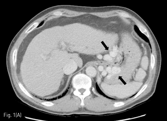
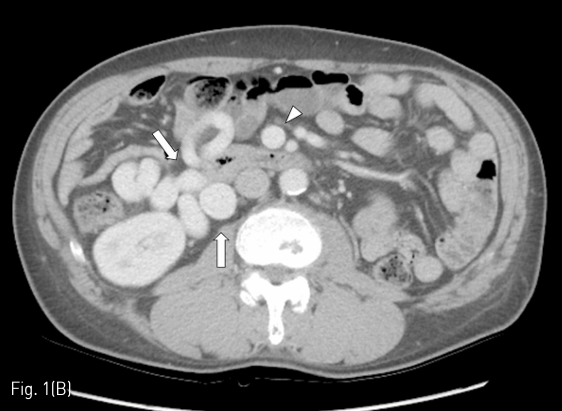
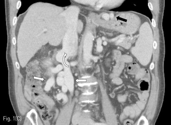
시술 전 시행한 dynamic liver CT에서 간경화 소견과 현저한 gastric varices, mesenteric-retroperitoneal varices가 관찰되었다 (Fig. 1A-C). Gastric varices는 gastro-renal shunt가, mesenteric-retroperitoneal varices는 meso-caval shunt가 각각의 경로로 생각되었다. Meso-caval shunt는 superior mesenteric vein (SMV)에서 right renal vein을 거쳐 inferior vena cava (IVC)로 유출되는 것으로 판단하였다. Esophageal varices도 보이긴 하였으나 경미한 정도였다.
시술방법 및 재료
환자의 right internal jugular vein을 초음파 유도하에 천자한 후 6-F sheath (Flexor Check-Flo sheath; Cook, Bloomington, IN, USA)를 삽입하고 0.035-inch, 180-cm-long hydrophilic guide wire (Terumo, Tokyo, Japan) 와 4-F angled-tip catheter (Cobra; Terumo)를 left renal vein을 통해 gastro-renal shunt로 진입시켰다. 이후 sheath를 shunt까지 진입시키고 16mm type II vascular plug (AGA medical, Golden valley, MN, USA)를 shunt의 가장 확장된 부위에 설치한 후 plug보다 근위부의 shunt까지 4-F angled-tip catheter를 진입시켰다. Shunt의 근위부까지 진입한 catheter를 유지한 채로 vascular plug를 shunt의 가장 좁은 부위까지 당겨 고정시킨 후 희석한 조영제와 gelatin sponges 혼합물을 catheter를 통해 주입하며 embolization을 시행하였다. 충분한 embolization을 시행한 이후에는 sheath를 통해 retrograde venography를 시행하여 shunt의 폐쇄를 확인한 후 catheter를 제거하고 vascular plug를 delivery cable에서 분리하였다 (Fig. 2A).
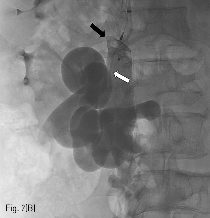
Gastro-renal shunt에 대한 embolization을 시행한 후, 다시 right internal jugular vein을 통해 7-F sheath (Flexor Check-Flo sheath; Cook, Bloomington, IN, USA)를 삽입하고 guide wire와 4-F angled-tip catheter를 right renal vein을 통해 meso-caval shunt로 진입시켰다. 이후 gastro-renal shunt와 같은 방법으로 20mm type II vascular plug (AGA medical, Golden valley, MN, USA)를 설치하고 4-F angled-tip catheter를 통해 gelatin sponges를 주입하며 embolization을 시행하였다. Embolization 이후 sheath를 통해 retrograde venography를 시행한 후 catheter를 제거하고 vascular plug를 delivery cable에서 분리하면서 시술을 완료하였다(Fig. 2B).
추적관찰
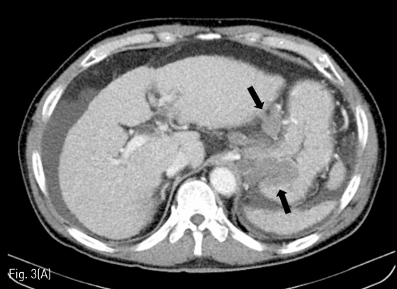
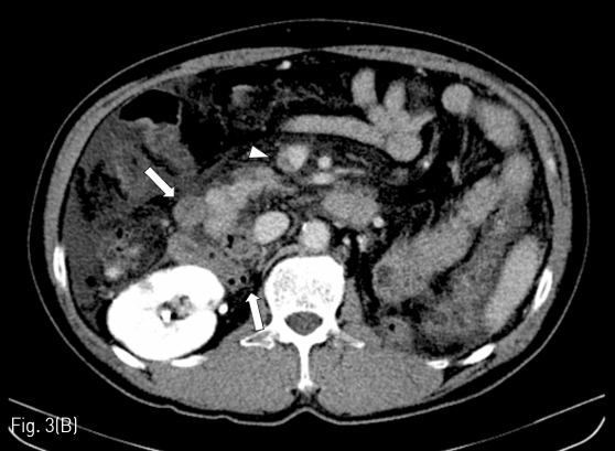
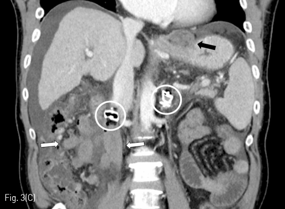
시술 2일 후 추적 검사로 시행한 dynamic liver CT에서 gastric and mesenteric-retroperitoneal varices의 complete thrombosis 소견을 확인할 수 있었다. 시술 후 CT에서 ascites가 새로 생겼으나 내과적으로 조절 가능한 수준이었다. 또한 SMV 내부에 partial thrombosis가 보였으나 임상적으로 문제가 되지는 않았다(Fig. 3A-C).
Fig 1A
(A-C) Contrast-enhanced CT images obtained before PARTO show gastric varices (black arrows) and mesenteric-retroperitoneal varices (white arrows). Arrow head indicates dilated SMV. Curved arrow indicates the meso-caval shunt.
Fig 1B
(A-C) Contrast-enhanced CT images obtained before PARTO show gastric varices (black arrows) and mesenteric-retroperitoneal varices (white arrows). Arrow head indicates dilated SMV. Curved arrow indicates the meso-caval shunt.
Fig 1C
(A-C) Contrast-enhanced CT images obtained before PARTO show gastric varices (black arrows) and mesenteric-retroperitoneal varices (white arrows). Arrow head indicates dilated SMV. Curved arrow indicates the meso-caval shunt.
Fig 2A
(A) After vascular plug (white arrow) placement in narrowest portion of the gastro-renal shunt via left inferior phrenic vein, additional embolization of the gastro-renal shunt, gastric varices, and afferent vein (arrow heads) was performed using gelatin sponges through the 4-F catheter (black arrow).
Fig 2B
(B) After vascular plug (black arrow) placement in narrowest portion of the meso-caval shunt, additional embolization of the meso-caval shunt and mesenteric-retroperitoneal varices was performed using gelatin sponges through the 4-F catheter (white arrow).
Fig 3A
(A-C) Contrast-enhanced CT images obtained 2 days after PARTO show complete thrombosis of gastric varices (black arrows) and mesenteric-retroperitoneal varices (white arrows). Arrow head indicates partial thrombosis of the SMV. White circles indicate plugs placed within the meso-caval shunt and the gastro-renal shunt.
Fig 3B
(A-C) Contrast-enhanced CT images obtained 2 days after PARTO show complete thrombosis of gastric varices (black arrows) and mesenteric-retroperitoneal varices (white arrows). Arrow head indicates partial thrombosis of the SMV. White circles indicate plugs placed within the meso-caval shunt and the gastro-renal shunt.
Fig 3C
(A-C) Contrast-enhanced CT images obtained 2 days after PARTO show complete thrombosis of gastric varices (black arrows) and mesenteric-retroperitoneal varices (white arrows). Arrow head indicates partial thrombosis of the SMV. White circles indicate plugs placed within the meso-caval shunt and the gastro-renal shunt.
고찰
Portal-systemic shunts(PSS)는 간경화 환자에서 간성 혼수를 유발할 수 있는 중요한 원인이다. PSS 중 가장 흔한 collateral pathway는 spleno-renal 혹은 gastro-renal shunt이며 superior mesenteric-caval shunt는 상대적으로 드물다. 1835년 Retzius가 portal venous system과 IVC 사이의 문합을 보고한 이후로 superior mesenteric-caval shunt는 veins of Retzius로 불리기도 한다. Veins of Retzius는 다음과 같이 4가지가 알려져 있다: (a) ileocolic vein이 right gonadal vein을 통해 IVC 혹은 right renal vein으로 배출되는 경로; (b) pancreatico-duodenal vein이 IVC로 배출되는 경로; (c) SMV의 분지가 left gonadal vein을 통해 left renal vein으로 배출되는 경로; (d) ileocolic vein이 직접 IVC로 배출되는 경로이다.
본 증례처럼 PSS를 동반한 간성 혼수 환자에서는 PSS를 차단하는 치료를 고려할 수 있으며 balloon-occluded retrograde transvenous obliteration (BRTO) 혹은 PARTO 같은 endovascular shunt occlusion이 비침습적이고 효과적인 치료로 널리 받아들여지고 있다. 일반적으로 gastro-renal shunt에 대 해 BRTO나 PARTO를 시행하는 경우가 가장 많으며 다른 PSS에 대한 치료를 시행하는 경우는 드물다. 저자들이 알기로는 meso-caval shunt를 BRTO로 치료한 증례 보고 들은 있으나 PARTO를 시행한 증례 보고는 없었다. 드물게 vascular plug를 사용한 증례 보고가 있기는 하지만 gelatin sponges를 사용하지 않고 coil을 사용하는 등 PARTO와는 다른 시술 방법으로 치료를 한 증례들이었다. 또한 본 증례처럼 gastro-renal shunt와 meso-caval shunt를 동시에 치료한 증례는 지금까지 보고된 바 없기에 본 증례를 보고하고자 하였다.
PARTO는 비교적 최근에 소개된 술식으로 balloon 과 sclerosants 대신 plug와 gelatin sponges를 이용하는 방법이다. BRTO에 비해 기술적으로 쉽고, 안전하며, 시술시간도 훨씬 짧다는 장점이 있다. Gastro-renal shunt의 치료에서 BRTO의 훌륭한 대안으로 주목을 받고 있는 시술 방법이며 때문에 다른 유형의 PSS에 대해서도 PARTO가 효과적일 것으로 기대할 수 있다. BRTO와는 달리 PARTO는 대부분의 경우 gastro-renal shunt의 측부 정맥에 대한 선택적 색전술을 시행하지 않아도 안전하게 시술을 시행할 수 있다. 하지만 meso-caval shunt를 폐색시키기 위해 PARTO를 시행할 경우에는 이론적으로 SMV나 portal vein의 thrombosis를 유발할 가능성이 있기 때문에 주의를 요한다. 아직 meso-caval shunt에서 PARTO를 시행한 데이터가 충분하진 않지만 BRTO를 시행한 증례 보고들에서는 SMV나 portal vein의 thrombosis가 보고된 바 있다. Mesenteric vein thrombosis의 경우에는 marginal vein 등 collateral pathway가 있으면 임상적으로 문제가 되지 않지만 시술 시 주의가 필요하다. Thrombosis가 발생했을 경우에는 oral antiplatelets이 치료에 효과적인 것으로 보고된 바 있다. 본 증례에서는 SMV에 partial thrombosis가 관찰되었으나 임상 증상은 유발하지 않는 경미한 정도였으며 특별한 치료는 하지 않았다.
BRTO와 마찬가지로 PARTO를 시행한 후 portal hypertension이 악화되어 ascites가 증가하거나 esophageal varices가 악화되는 등의 부작용이 있을 수 있다. 본 증례에서도 CT 추적 검사에서 ascites가 새로 관찰되었으나 내과적으로 조절 가능한 정도였다. Esophageal varix는 추적 관찰 기간이 짧아 CT로 평가하기에는 제한이 있었으며 내시경 추적 검사는 아직 시행하지 않았다.
본 증례는 추적 관찰 기간이 짧은 단일 증례이지만 meso-caval shunt에서도 PARTO가 안전하고 효과적으로 적용될 수 있음을 보여주는 증례로서 의의가 있다. 향후 이러한 경험이 축적되면 PARTO가 보다 다양한 적응증에 적용될 수 있을 것으로 기대된다.
참고문헌
1. Ibukuro K, Tsukiyama T, Mori K, Inoue Y. Veins of Retzius at CT during arterial portography: anatomy and clinical importance. Radiology1998;209:793-800
2. Boixadera H, Tomasello A, Quiroga S, Cordoba J, Perez M, Segarra A. Successful embolization of a spontaneous mesocaval shunt using the Amplatzer Vascular Plug II. Cardiovasc Intervent Radiol 2010;33:1044-1048
3. de Martinis L, Groppelli G, Corti R, et al. Disabling portosystemic encephalopathy in a non-cirrhotic patient: Successful endovascular treatment of a giant inferior mesenteric-caval shunt via the left internal iliac vein. World J Gastroenterol 2017;23:8426-8431
4. Gwon DI, Ko GY, Yoon HK, et al. Gastric varices and hepatic encephalopathy: treatment with vascular plug and gelatin sponge-assisted retrograde transvenous obliteration--a primary report. Radiology 2013;268:281-287
5. Minamiguchi H, Kawai N, Sato M, et al. Balloon occlusion retrograde transvenous obliteration for inferior mesenteric vein-systemic shunt. J Vasc Interv Radiol 2011;22:1039-1044
6. Ibukuro K, Sugihara T, Tanaka R, et al. Balloon occluded retrograde transvenous obliteration (BRTO) for a direct shunt between the inferior mesenteric vein and the inferior vena cava in a patient with hepatic encephalopathy. J Vasc Interv Radiol 2007;18:121-125
Citations
Citations to this article as recorded by